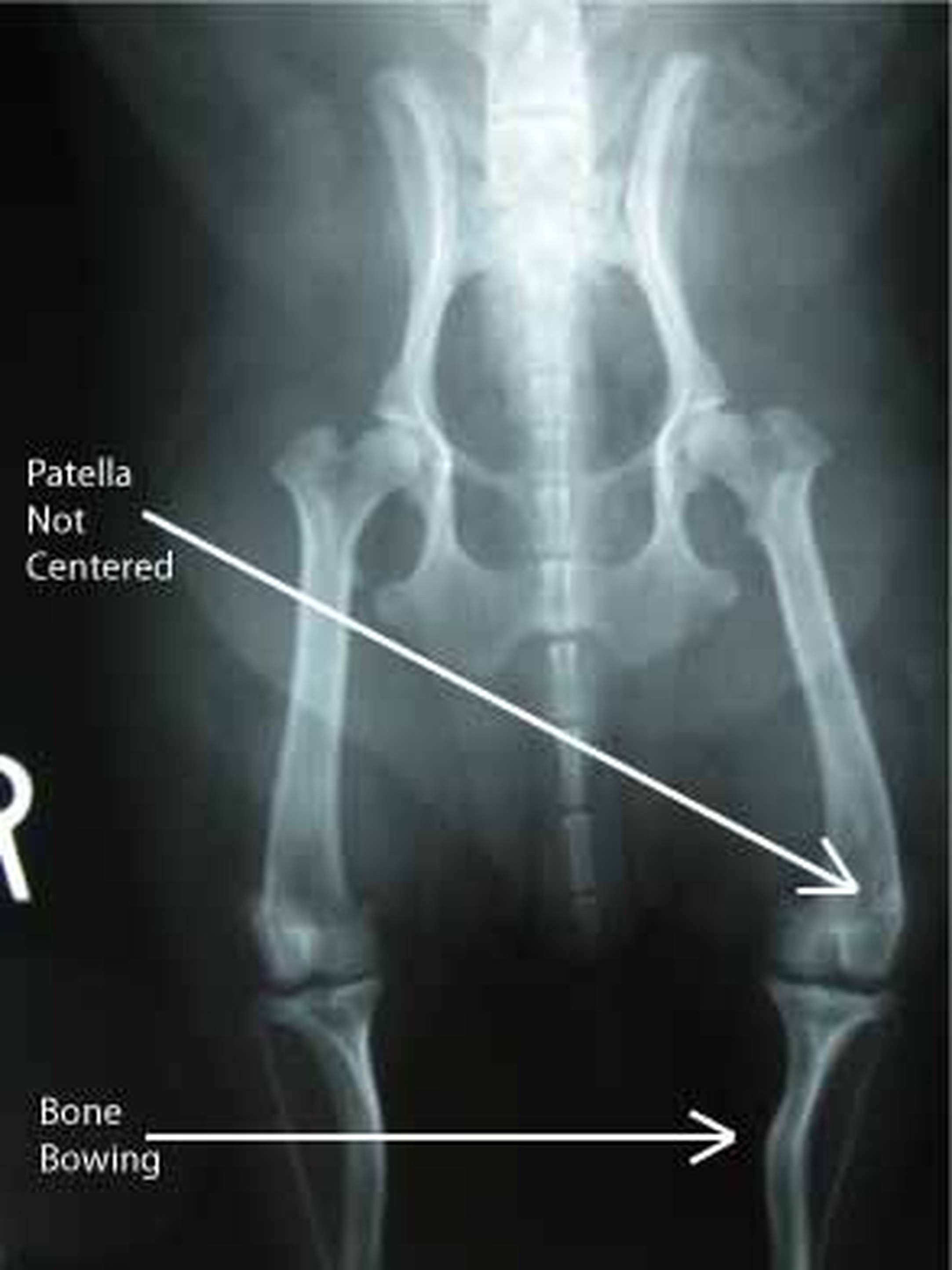
 | Patellar Luxation in Dogs Patellar luxation occurs when the dog's kneecap (patella) is dislocated from its normalanatomic position in the groove of the thigh bone (femur). When the kneecap is dislocated from the groove of the thigh bone, it can only be returned to its normal position once the quadriceps muscles in the hind legs of the animal relax and lengthen. It is for this reason that most dogs with the condition will hold up their hind legs for a few minutes. A dislocated kneecap is one of the most prevalent knee joint abnormalities in dogs. The condition is most common in toy and miniature dog breeds such as the Yorkshire Terrier, Dachshund, West Highland White Terrier (Westie), Pomeranian, Pekingese, Chihuahua, and Boston Terrier. Female dogs are 1 1/2 times more likely to acquire the condition. Symptoms and Types The specific symptoms of a dislocated kneecap will depend on the severity and persistence of the condition, as well as the amount of degenerative arthritis that is involved. Typically, a dog with a dislocated kneecap will exhibit prolonged abnormal hindlimb movement, occasional skipping or hindlimb lameness, and sudden lameness. The dog will rarely feel pain or discomfort once the kneecap is out of position, only feeling pain at the moment the kneecap slides out of the thigh bone's ridges. Causes A dislocated kneecap is usually caused by a genetic malformation or trauma. The clinical signs of the condition will normally start showing approximately four months after birth. Diagnosis A dislocated kneecap is diagnosed through a variety of means. Top view (craniocaudal) and side view (mediolateral) X-rays of the stifle joint, hip, and hock may be used to detect bending and twisting of the thigh bone and larger bone of the lower leg. Skyline X-rays may reveal a shallow, flattened, or curved groove of the thigh bone. A fluid sample taken from the joint and an analysis of the lubricating fluid in the joint (synovial fluid) will show a small increase in mononuclear cells. It is also necessary for the veterinarian to perform an examination by touch to feel for kneecap freedom. Treatment Medical treatment for kneecap dislocation has very little effectiveness; surgery is the preferred treatment of choice for severe cases. Surgery can correct both the affected structures and the movement of the kneecap itself, and in 90 percent of cases, frees the dog from lameness and dysfunction. The kneecap may be fastened on the outside of the bone to prevent it from sliding towards the inside. Alternatively, the groove of the thigh bone may be deepened so that it can better hold the kneecap. Living and Management Follow-up treatment after successful surgery will include leash walk exercise for one month (avoid jumping) and yearly examinations to check for progress. It is important that pet owners are aware that there is a high possibility of recurrence (48 percent), although the dislocation will be considerably less severe than the original incidence. Because kneecap dislocation is genetically inherited, the breeding of affected dogs is highly discouraged. PreventionThere are currently no known preventative measures for this medical condition. |
|
1 Comment
Urolithiasis, Calcium Oxalate in Cats
Urolithiasis is described as the presence of stones in the urinary tract. When these stones are made of calcium oxalate, they are referred to as calcium deposits. In most cases the stones can be removed safely, giving the cat a positive prognosis. Symptoms and Types Although it is rare in cats, the most common symptom of urolithiasis is straining while the animal is urinating. If there is inflammation in the urinary tract, the cat may have an enlarged belly or the area surrounding the urinary region may be noticeably irritated. If the calcium deposits are large, they can sometimes be felt through the skin by a veterinarian. Causes The primary cause for the formation of stones is high levels of calcium in the urine. Some risk factors can include excessive dietary protein or vitamin D, a vitamin B6 deficiency, the use of calcium supplements or steroids, and a diet comprised exclusively of dry food. The most common breeds to develop the medical condition include the Himalayan, the Scottish Fold, the Persian, the Ragdoll, and the Burmese. Diagnosis X-rays and ultrasounds are performed to rule out any other underlying medical conditions which may be causing the cat's pain or trouble urinating. Blood work will be done to examine the cat's nutrient levels and determine if any are outside of the normal range. Treatment One of the most common treatment options is the surgical removal of the stones; in some cases shock waves can be used to help break up the stones. Also, depending on the size and severity of the stones, they can occasionally be flushed and massaged out of the cat's system with a catheter and fluids. Living and Management It is important to reduce the cat's activity levels following surgery. Possible complications from the formation of these stones may arise such as the blockage of the urinary tract and the cat's inability to urinate. It is common for animals to reform these calcium-based stones over time. Treatment on an ongoing basis will include the monitoring of calcium intake and the urinary patterns of the cat to observe if any problems develop. If surgery was used to remove the stones, post-surgical X-rays are recommended to ensure that the stones were completely removed. Prevention The best prevention of recurrence is to monitor the cat's calcium levels on an ongoing basis so that adjustments can be made in the diet to maintain normal calcium levels. Generalized Tremor Syndrome in Dogs
Shaker syndrome is a disorder which causes a dog's entire body to shake. It is also known as idiopathic cerebellitis, which describes inflammation of the cerebellum (the part of the brain that is responsible for the coordination and regulation of voluntary muscular movement) for unknown reasons. While dogs of any coat color can be affected, those with a white hair coat are over-represented in the medical literature. For example, Maltese and West Highland White Terrier (Westie) appear to be predisposed. In addition, both genders are affected by shaker syndrome, especially young to middle-aged dogs. Symptoms and Types
Causes Although a dog may be affected by the syndrome due to unknown reasons (idiopathic), it is most often associated with mild central nervous system disease. Diagnosis You will need to provide a thorough history of your dog's physical and behavioral health leading up to the onset of symptoms. Your veterinarian will perform a complete physical exam on your dog, including standard laboratory work, such as a blood chemical profile, a complete blood count, a urinalysis and an electrolyte panel to rule out other diseases. A cerebrospinal fluid (fluid from the spinal cord) sample may also be taken by your veterinarian and sent to the laboratory for analysis of the nervous system. Your doctor will use the process of differential diagnosis to rule out each of the more common causes until the correct disorder is settled upon and can be treated appropriately. Some other causes for the tremors can be anxiety/fear, seizures, and hypothermia. Treatment Depending on how severe the tremors are, and your dog's overall condition, care will be given inpatient or outpatient. If your dog is very ill as the result of tremors, or if there is an underlying condition or infection, your dog will be hospitalized until its health stabilizes. The primary treatment for neurological shaker syndrome is the use of corticosteroids for reducing the inflammatory response in the body. Most dogs recover in a week although some rare patients never entirely recover. The steroids will be gradually reduced over the course of a few months until they are not being used anymore. Steroid treatment will be reinstated if symptoms recur, and in some cases, steroid treatment will need to be continued for a longer period and possible even the lifetime of the dog in order to maintain health. Living and Management Your veterinarian will schedule weekly evaluations for your dog for the first month after the initial treatment. Thereafter, your veterinarian will schedule monthly follow-up appointments with you for your pet until the corticosteroids are discontinued. |
PET SCAN Blog:
The INNER Pet... Pet Health Inside & Out The goal of this blog is to help educate pet owners by sharing pet health facts and interesting pet news articles Archives
September 2019
Categories
All
|
 RSS Feed