Pet Scan:  Mobile Veterinary Ultrasound Service
  • Home
  • Indications for Ultrasound
  • Services
  • What To Expect
  • Our Doctors
  • Referring DVMs

New Research May Help Control Brown Dog Ticks

6/8/2015

0 Comments

 
New research may lead to more effective ways to control the brown dog tick, a species that can cause major problems for dogs and their owners.

University of Florida scientists say they found that the brown dog tick is resistant to permethrin, a widely used anti-tick chemical. They also found that carbon dioxide seems to be an effective way to lure ticks to bug traps.

Unlike other ticks, the brown dog tick can complete its life cycle indoors. One female brown dog tick can lay up to 5,000 eggs in its lifetime, the researchers said.

The bugs hide in hard-to-reach places. Some dog owners take desperate steps to be free of these ticks. These steps may include giving away their dogs, fumigating their homes, throwing out many possessions, or even moving, the researchers said.

"They're particularly troublesome for people who have cluttered homes, and they drive some homeowners to desperate measures in search of ways to control the tick," Phil Kaufman, an associate professor of veterinary entomology at the University of Florida, Gainesville, said in a university news release. "Eliminating places where ticks live and breed is one of the best practices for tick control."

Because the researchers found that the ticks are resistant to permethrin, pet owners and pest control companies should use the chemical fipronil. This anti-tick chemical should work in most cases, the researchers said.

However, dog owners should watch for loss of effectiveness with fipronil. An indication that fipronil isn't working is seeing ticks that appear to be alive and swelling within the month after treatment, the researchers said.

In addition to using pesticides, the researchers said vacuuming can help control ticks, too.

The finding on carbon dioxide suggests it may be possible to lure ticks from their hiding spots in nooks and crannies throughout the house to one location. This makes it easier to control them, according to Kaufman.

The results were published recently in the Journal of Medical Entomology.

Source: University of Florida, news release, May 19, 2015 / Robert Preidt

0 Comments

Valley Fever (Coccidioidomycosis)

4/16/2015

0 Comments

 
San Joaquin Valley Fever, now called Valley Fever for short, was named because of an outbreak among farm workers in the well known San Joaquin Valley of California. The condition is caused by inhaling dust or dirt contaminated with a fungus called Coccidioides immitis. The fungus is inhaled into the lungs where it may simply cause a low grade chronic cough and calcification (mineralization) of local lymph nodes or, if the immune system is weak, it can cause pneumonia or even disseminate throughout the body.

About the Fungus

The fungus lives only in one area: the Lower Sonoran life zone. This area includes the southwest United States, Mexico and Central America as well as parts of South America. In these places, soil is sandy and alkaline. There is little rainfall. Summer is hot and winter is cold. Fungal spores, called arthroconidia, are released from the ground after a rainfall and are carried by the wind. Earthquakes or high winds or even crop harvesting have led to outbreaks of this infection as both conditions stir up fungal spores. Once the spores are in the air, they can be inhaled and set up infection.

Getting Sick

It has been estimated that 60% of animals and people that become infected with this fungus never get sick. It is only when antibodies indicating past infection show up on a blood test that infection is discovered.

But the other 40% do get sick. It only takes 1 to 3 weeks from the inhalation of fungal spores before coughing starts. Fever, weight loss, and appetite loss accompany the cough, which can be dry or productive. If the fungus escapes the lung and travels out elsewhere in the body, the infection has disseminated and prognosis is much worse. On the average, several months pass between the initial cough (which may be so mild as to have gone undetected) and signs in another organ system. There is an order by which the fungus disseminates in that it appears in certain organs before others. After the lung, the fungus spreads to bone, then eyes, heart, testicles, brain, spinal cord, and abdominal organs. In cats, the pattern is a bit different with the usual disease picture being draining skin lesions with fever, weight loss and poor appetite. The respiratory phase is rarely noted in feline patients.

An animal or person who is sick is not contagious; infection is only through inhalation of a fungal spore released from dirt, but only a few spores are necessary for infection to occur.

Testing

There are several diagnostics that can be helpful with this condition.

  • Radiographs
    The patient with a cough is likely to have x-rays taken of the chest. Findings will not be specific to Valley Fever; large lymph nodes and lung infiltration will likely be found. The point is to rule out other conditions that are more readily identifiable on radiographs such as heart disease, tracheal collapse, or more localized forms of pneumonia.
  • Cytology
    If some sort of tracheal wash or lavage is performed, where samples of lung fluid are collected for analysis, it is possible to capture some fungal spores. If this occurs, the diagnosis is confirmed, as Coccidioides immitis has a fairly characteristic large size and appearance.


  • Blood testing
    Blood testing is by far the least invasive way of diagnosing Valley Fever. Antibodies against the fungus are measured in a blood sample using one or even several different methods (tube precipitin, complement fixation, latex agglutination, gel immunodiffusion, or enzyme linked immunoassay). As mentioned, many patients are exposed to this fungus and do not get sick. These patients will absolutely have positive antibody tests so it is important not to treat an animal with a positive antibody level (also called an antibody titer) and no history of symptoms of any kind.
Treatment

A general rule with any fungal infection is that it takes a long time to clear. Valley Fever is no different and a year of medication administration is fairly common. Treatment continues until the clinical signs have resolved, the radiographs look normal, and the antibody level has stabilized (note antibody levels may never fully drop to zero). Ketoconazole, itraconazole,  and fluconazole are the usual drugs to treat this condition. Monitoring liver enzymes is important with all of these, particularly since long treatment courses are common.

Lufenuron, the active ingredient in the flea control product Program, was developed to inhibit the development of chitin (the crunchy exterior material of the insect body). The idea was that a flea larva who had fed upon blood pellets (flea dirt) from a pet treated with oral lufenuron would be unable to properly pupate into an adult flea. It turns out that the shell of Coccidioides immitis is also rich in chitin and that lufenuron at specific doses may be helpful in clearing this infection. This is still an experimental use but it may be worthwhile to ask your veterinarian about this product.

If the disease is limited to the lung, prognosis is felt to be good. The most severe form of infection appears to involve bone and when multiple bones are infected, complete recovery is unlikely unless amputation is possible. Eye involvement is painful and very difficult to clear short of removing the eye (enucleation). Obviously, a patient with any internal fungal infection should avoid immunosuppressive drugs for other conditions as their use could lead to a disastrous fungal dissemination.

Source: Veterinary Partner

0 Comments

Immune Mediated Hemolytic Anemia (IMHA)

3/20/2015

1 Comment

 
Immune-mediated hemolytic anemia is a condition where the patient’s immune system begins attacking her own red blood cells. What occurs on a microscopic level is that the branch of the immune system that produces antibodies begins to direct them against the patient’s own red blood cells. The red blood cells quickly become coated with tiny antibody proteins, essentially marking these red blood cells for destruction. When too many red blood cells are destroyed the patient is said to be anemic and will feel cold and weak. Because the red blood cells are being destroyed internally (and not lost through bleeding), the patient will become yellow-tinged (jaundiced) rather than pale (see picture below).

Removing old Red Cells/Recycling Iron: The Normal System

Red blood cells have a natural life span from the time they are released from the bone marrow to the end of their oxygen-carrying days when they become too stiff to move through the body’s narrow capillaries. A red blood cell must be supple and flexible to participate in oxygen delivery and carbon dioxide removal, so when the cell is no longer functional the body destroys it and recycles its components.

When old red blood cells circulate through the spleen, liver, and bone marrow, they are plucked from circulation and destroyed in a process called extravascular hemolysis. Their iron is sent to the liver for recycling in the form of a yellow pigment called bilirubin. The proteins inside the cell are broken down into amino acids and used for any number of things (burning as fuel, building new protein etc.) The spleen uses immunological cues on the surface of red blood cells to determine which cells are plucked out of circulation. In this way, red cells parasitized by infectious agents are also removed from circulation along with the geriatric red cells. When the immune system marks too many cells for removal, problems begin.

The jaundiced/icteric pet has a yellow color in the gums and whites of the eyes.The spleen enlarges as it finds itself processing far more damaged red blood cells than it normally does. The liver is overwhelmed by large amounts of bilirubin and the patient becomes jaundiced (icteric), which means her tissues become a yellow/orange color.

Making matters worse, a protein system called the complement system is activated by these anti-red cell antibodies. Complement proteins are able to simply rupture red blood cells if they are adequately coated with antibodies, a process called intravascular hemolysis. Ultimately, there aren’t enough red blood cells left circulating to bring adequate oxygen to the tissues and remove waste gases.  A life-threatening crisis has emerged; in fact 20-80% mortality (depending on the study) has been reported with this disease.

Signs you Notice at Home
 
The jaundiced/icteric pet has a yellow color in the gums and whites of the eyes. Illustration by Wendy Brooks.Your pet is obviously weak. She has no energy and has lost interest in food. Urine is dark orange or maybe even brown. The gums are pale or even yellow-tinged as are the whites of the eyes. There may be a fever. You (hopefully) brought your pet to the veterinarian’s office as soon as it was clear that there was something wrong.
 
The Tests Show Anemia

Part of a general evaluation includes blood testing. If your pet seemed obviously pale or jaundiced (yellow-tinged), right off the bat your vet may run a test called a packed cell volume. This test can be run in most veterinary clinics and involves spinning a small amount of blood in a small glass tube at high speed to separate the red blood cells from the serum (the blood’s liquid phase). By comparing the blood tube to a chart, it will become obvious if your pet has a low red blood cell count. If your pet has hemolytic anemia, the serum will be bright orange instead of its normal off-white color.

The anemia may be seen on a full blood panel sent to a reference laboratory. These laboratories perform a test called a hematocrit, which is slightly different from a packed cell volume but essentially measures the same thing: the percentage of blood volume made up by red blood cells. This number should be 43 - 59 for dogs and 29 - 50 for cats.

Anemia is the condition where one’s red blood cell count is low. Anemia can be mild or severe and can represent bleeding, red blood cell destruction (as in IMHA), or simply lack of red blood cell production. Once a patient is found to be anemic, it is important to determine why.

The Tests Show Responsive Anemia

Anemia due to poor red blood cell production by the bone marrow is called a non-responsive anemia. Such anemias are caused by chronic inflammatory diseases (like inflamed skin, infected teeth, or other long standing irritations), kidney failure, cancers of various types, or certain drugs (especially agents of chemotherapy).

Normally when red blood cells are lost, the drop in blood oxygen that results causes hormonal changes leading to increased production of red blood cells by the bone marrow. These are called responsive anemias because the bone marrow is responding. Bleeding and immune mediated red blood cell destruction are both responsive anemias.

There are several ways to determine if the anemia is responsive or not from the blood panel results. Most blood panels run by reference laboratories include a portion called a complete blood count, or CBC, which reviews red blood cell count, size, shape, and maturity as well as white blood cell types and ratios. A patient with a responsive anemia will have an active bone marrow. Red blood cells will be released early leading to a variety of sizes and redness of red blood cells circulating in the blood (less mature red blood cells are larger and paler than mature cells). Further, red blood cell precursors called reticulocytes are released. (These may be thought of as red blood cells so immature they can’t truly be called red blood cells yet.) If the bone marrow stimulus is especially strong, red blood cells may be released still containing cell nuclei.

These findings indicate the anemia is responsive. This means either red blood cells are being lost through bleeding, possibly internal bleeding, or they are being destroyed by the immune system.

Which is it?

The Tests Suggest Immune-Mediated Destruction rather than Bleeding

There are several clues in blood testing that tell us if our patient is bleeding or destroying red blood cells. You might think it would be obvious if the patient were bleeding but if the bleeding is internal it may not be so obvious.

Icterus (also Called Jaundice)
We have mentioned this but let’s recap. Icterus is the yellow color that is taken up by a patient’s tissues when the liver is overwhelmed with bilirubin, the iron-containing by-product of red blood cell destruction.  Normally red blood cells are removed from the circulation when they become old and inflexible. Their iron is recycled in the liver. With so many red cells being destroyed, the liver is overwhelmed and bilirubin (a yellow pigment) spills out everywhere, coloring urine, gums, skin, and the eyes orange.

Is immune-mediated red cell destruction the only cause of icterus? Absolutely not. Liver failure also leads to icterus when the diseased liver cannot process normal amounts of bilirubin. In cats especially, bacterial endotoxin (the toxic cell walls of certain types of infecting bacteria) can lead to icterus. Usually, however, a responsive anemia together with icterus, suggests immune-mediated red cell destruction.

Spherocytes

A normal red blood cell is concave on both sides and shaped like a disc. It is slightly paler in the middle than on its rim. After a portion has been bitten off, it re-shapes into a more spherical shape with a denser red color. The presence of spherocytes indicates that red blood cells are being destroyed.

Autoagglutination
In severe cases of immune-mediated hemolytic anemia, the immune destruction of red cells is so blatant that the red cells clump together (because their antibody coatings stick together) when a drop of blood is placed on a microscope slide. Imagine a drop of blood forming not a red spot but a yellow spot with a small red clump inside it. This finding is especially foreboding.

Leukemoid Reaction
Classically, in IMHA the stimulation of the bone marrow is so strong that even the white blood cells lines (which have little to do with this disease but which also are born and incubate in the bone marrow alongside the red blood cells) are stimulated. This leads to white blood cell counts that are spectacularly high.

More Tests Needed

Coomb’s Test (also Called a Direct Antibody Test)
This test is designed to identify the antibodies that are coating red blood cell surfaces. This test is the current state of the art for the diagnosis of IMHA but, unfortunately, it is not as helpful as it might seem. It can be erroneously positive if there is any inflammation or infectious disease (which might lead to harmless attachment of antibody to red cell surfaces) or in the event of a prior blood transfusion (ultimately transfused red cells are removed from the immune system). The Coomb’s test can be erroneously negative for a number of reasons as well. If the clinical picture fits with IMHA, often the Coomb’s test is skipped.

Remember, not all causes of hemolysis (red blood cell destruction) are immune-mediated.  Onions in large amounts, and possibly garlic, will cause a toxic hemolysis. Zinc toxicity, usually caused from swallowing a penny minted after 1983 or from licking a zinc oxide ointment applied to the skin, will cause hemolysis as well. In a young animal, a genetic red blood cell malformation might be suspected.

Once there is a diagnosis of IMHA, efforts to determine an underlying cause should be made. 

Treatment and Monitoring During the Crisis

The patient with IMHA is often unstable. If the hematocrit has dropped to a dangerously low level, then blood transfusion is needed. It is not unusual for a severely affected patient to require many transfusions. General supportive care is needed to maintain the patient’s fluid balance and nutritional needs. Most importantly, the hemolysis must be stopped by suppressing the immune system’s rampant red blood cell destruction. We will review these aspects of therapy.

Transfusion
There are several products that may be helpful in treating the IMHA case. If the patient is in a crisis and needs immediate therapy, artificial blood may be a good choice. Artificial blood (Oxyglobin®) is made from hemoglobin harvested from cow’s blood. Because the patient does not receive actual red blood cells, the artificial blood does not further stimulate the immune system. Artificial blood does not require refrigeration and is likely sitting on the shelf ready to use at your veterinarian’s office. The disadvantage of artificial blood is that it does not last in the body like a well-matched blood transfusion does. The body begins removing artificial blood immediately so that the entire transfusion is probably gone in 48 hours or so. In IMHA, this may buy some time but since IMHA tends to have a long treatment course, it is likely that the patient will be back where they started from at that point. If a compatible donor is not readily available, sometimes an artificial blood transfusion buys enough time to find a compatible donor

Well-matched whole blood or packed red cells (a unit of whole blood with most of the plasma, leaving only a concentrated solution of red blood cells) may last longer. Compatible blood can last a good 3 to 4 weeks in the recipient’s body. The problem, of course, with IMHA is that even the patient’s own red blood cells are being destroyed so what chance do donated cells have? Cross matching of red cells is ideal but still may not lead to a good match given the hyperactivity of the patient’s immune response. For this reason, it is not unusual for several transfusions to become necessary during treatment.

Immune Suppression
Corticosteroid hormones in high doses are the cornerstone of immune suppression. Prednisone and dexamethasone are the most popular medications selected. These hormones are directly toxic to lymphocytes, the cells that produce antibodies. If the patient’s red blood cells are not coated with antibodies, they will not have been targeted for removal so stopping antibody production is an important part of therapy. These hormones also suppress the activity of the reticuloendothelial cells that are responsible for removing antibody-coated red cells.

Corticosteroids may well be the only immune suppressive medications the patient needs. The problem is that if they are withdrawn too soon, the hemolysis will begin all over again. The patient is likely to be on high doses of corticosteroids for weeks or months before the dose is tapered down and there will be regular monitoring blood tests. Expect your pet to require steroid therapy for some 4 months; many patients must always be on a low dose to prevent recurrence.

Corticosteroids in high doses produce excessive thirst, re-distribution of body fat, thin skin, panting, predisposition for urinary tract infection and other signs that constitute Cushing’s Syndrome. This is an unfortunate consequence of long-term steroid use but in the case of IMHA, there is no way around it. It is important to remember that the undesirable steroid effects will diminish as the dosage diminishes.

More Immune Suppression

If no response at all is seen with corticosteroids, supplementation with stronger immune suppressive agents is necessary. The two most common medications used in this case areazathioprine and cyclophosphamide. These are serious drugs reserved for serious diseases. Follow the links above to read more about specific side effects, concerns, etc.

Cyclosporine is an immune-modulator made popular in organ transplantation technology. It has the advantage over the two above medications of not being suppressive to the bone marrow cells. It has been a promising adjunctive therapy in IMHA but has two major problems: first, it is extremely expensive and second, blood level monitoring is necessary to ensure that the dosage is appropriate. This adds dramatically to the expense of treatment but ultimately may provide results not possible with other drugs.

Leflunomide is an immuno-modulator that is meant for patients with immune mediated diseases when corticosteroids either do not work or cannot be used. It is expensive (approximately $600 per month) but we may be hearing more about it in the future.

Human gamma globulin transfusion is a treatment that is reserved for patients for whom more traditional methods of immune suppression have been ineffective. The gamma globulin portion of blood proteins includes circulating antibodies. These antibodies bind the reticulo-endothelial cell receptors that would normally bind antibody-coated red blood cells. This prevents the antibody-coated red blood cells from being removed from the circulation. Human gamma globulin therapy seems to improve short-term survival in a crisis but, unfortunately, its availability is limited and it is very expensive.

Why did this Happen to your Pet?

When something as threatening as a major disease emerges, it is natural to ask why it occurred. Unfortunately, if the patient is a dog, there is a good chance that there will be no answer to this question. Depending on which study you read, 60-75% of IMHA cases do not have apparent causes.

In some cases, though, there is an underlying problem: something that triggered the reaction. A drug can induce a reaction that stimulates the immune system and ultimately mimics some sort of red blood cell membrane protein. Not only will the immune system seek the drug but it will seek proteins that closely resemble the drug and innocent red blood cells will be consequently destroyed. Drugs most commonly implicated include penicillins, trimethoprim-sulfa, and methimazole.

Drugs are not the only such stimuli; cancers can stimulate exactly the same reaction (especially hemangiosarcoma).

Red blood cell parasites create a similar situation except the immune system is aiming to destroy infected red blood cells. The problem is that it gets over-stimulated and begins attacking the normal cells as well.

There is some thinking that vaccination can trigger IMHA. Insect bites have also been implicated. Both have been temporally associated with the development of autoimmune hemolytic anemia. The relationship between recent vaccination and IMHA development is one of the factors that has led most universities to vaccinate dogs with the standard DHLPP every three years rather than annually.

Dog breeds predisposed to the development of IMHA include: cocker spaniels, poodles, Old English Sheepdogs, and Irish setters.

In cats, IMHA generally has one of two origins: feline leukemia virus infection or infection with a red blood cell parasite called Mycoplasma hemofelis (previously known as Hemobartonella felis).

Complications of IMHA

Thromboembolic Disease
This particular complication is the leading cause of death for dogs with IMHA (between 30-80% of dogs that die of IMHA do so due to thromboembolic disease). A thrombus is a large blood clot that obstructs (occludes) a blood vessel. The vessel is said to be thrombosed. Embolism refers to smaller blood clots spitting off the surface of a larger thrombus. These mini-clots travel and obstruct smaller vessels, thus interfering with circulation. The inflammatory reaction that normally ensues to dissolve errant blood clots can be disastrous if the embolic events are occurring throughout the body.

Heparin, a natural anticoagulant, may be used as a preventive in hospitalized patients or in patients with predisposing factors for embolism.

*Four to seven days are required for the bone marrow to generate a response. If hemolysis occurs suddenly there may not have been adequate time for a response. When this occurs, if there is any question about the responsive nature of the anemia, continued monitoring of the complete blood count will show a clear response in an appropriate time period. It should also be noted that in an especially unlucky patient, the red blood cell destruction may extend to the pre-red blood cells (reticulocytes, nucleated red cells and other precursors) within the bone marrow. If these cells are also destroyed, the condition is especially dangerous and it will take weeks rather than days to begin to see a response to treatment. The lack of circulating immature red cells will lead this anemia to test as non-responsive.

The 2002 study by Drs. Anthony Carr, David Panciera, and Linda Kidd at the University of Wisconsin School of Veterinary Medicine reviewed 72 dogs with IMHA looking for trends. Their findings are:

  • The only predisposed breed they found was the cocker spaniel.  
  • Most patients were female.  
  • The mean age was 6.8 years.  
  • Timing of vaccination was not associated with the development of IMHA.  
  • 94% of cases had spherocytes on their blood smears. 
  • 42% showed autoagglutination.  
  • 70% also had low platelet counts.  
  • 77% were Direct Coombs' positive.  
  • 58% were suspected of having disseminated intravascular coagulation.  
  • 55% required at least one blood transfusion.  
  • Mortality rate was 58%.  
  • Of those that died, 80% had thromboembolism present on necropsy (autopsy).

Prognostic Factors for Mortality and Thromboembolism in Canine Immune-Mediated Hemolytic Anemia. A.P. Carr, D. Panciera, L. Kidd. Journal of Veterinary Internal Medicine. 2002; 16: 504-509.

Another study
The 2005 study looking for trends by Drs. Tristan Weinkle, Sharon Center, John Randolph, Stephen Barr, and Hollis Erb at Cornell University reviewed 151 dogs with IMHA. They found:

  • Cockers spaniels and miniature schnauzers were both overrepresented (i.e., felt to be predisposed). These breeds, however, showed the same mortality rate as other breeds.  
  • Unspayed female dogs were overrepresented.  
  • Neutered male dogs were more commonly affected than unneutered male dogs (begging the question of whether male hormones might have some protective effect).  
  • The chance of survival either long term or short term was significantly enhanced by the addition of aspirin to the treatment protocol, especially when combined with azathioprine.  
  • Adequate vaccination information was not obtained for enough patients to comment on association with vaccination.  
  • 89% of affected dogs showed spherocytes on their blood smears.  
  • 78% showed autoagglutination.  
  • 70% of patients required at least one blood transfusion.  
  • Of the 151 dogs studied, 76% survived, 9% died, and 15% were euthanized. Survivors were hospitalized an average of 6 days. Non-survivors were hospitalized an average of 4 days.  
  • 100% of dogs that died or were euthanized showed thromboembolism on necropsy (autopsy).  
  • Of the dogs that survived 60 days or more, 15% experienced relapse. Most dogs treated with corticosteroids, azathioprine, and ultra-low dose aspirin did not experience relapse.

Source: Veterinary Partner
1 Comment

Using Viruses to Treat Cancer in Pets

12/12/2014

1 Comment

 
Surgery, radiation, and chemotherapy are the more commonly known treatments for cancer in pets. But newer technologies are opening up other possibilities. A recent experiment summary (abstract) suggests the use of genetic modified viruses to treat various types of cancer.

Oncolytic Virotherapy
The idea of using a virus for cancer treatment or oncolytic virotherapy is not a new idea. In the 1940s scientists conducted animal studies using viruses to treat tumors. Doctors in the 1950s observed that cancer patients that were stricken with viral infections or recently vaccinated experienced improvement in their condition. It was believed that the infections or vaccinations triggered an immune response that increased the production of interferon and tumor necrosis factors, or TNFs.

Interferons are large molecules released by cells infected with viruses, bacteria, parasites and tumors to interfere, hence their name, with virus reproduction and to trigger responses from immune cells. Interferons activate natural killer white blood cells and large white cells called macrophages that attack and destroy the invading organisms and cancer cells. Interferon promotes the production or molecular complexes that attach to viral, bacterial, parasite, and tumor cells so they are more quickly and effectively attacked by killer white cells. TNFs causes destructive changes in cell walls and causes foreign or tumor cells to burst and die

Despite the potential for viral therapy of cancer in these early years, it required the present advances in technology to achieve a real possibility. Precisely, it required our present ability to genetically modify organisms like viruses and safely use them to target cancer cells. The viruses are modified to prevent their normal ability to cause disease and genetically altered to produce interferon or other anti-cancer molecules.

Preliminary Study in Dogs
The abstract was based on a small study intended to evaluate the safety and effectiveness of a new oncolytic virus. The group was made up of of seven dogs suffering from various cancers (lymphoma, malignant melanoma and multiple myeloma). The researchers used a novel virus for their study; they used a modified vesicular stomatitis virus that causes oral, udder, and hoof ulcers in cattle. Although seldom fatal, the disease causes inappetence and decreased milk or meat production [in cattle]. It can also infect horses and pigs, and rarely, sheep, goats, and llamas. Because of its effect on agricultural production, vesicular stomatitis is a diagnosis that requires mandatory reporting to federal and state animal health officials.

The virus was also modified to produce human or canine interferon. Three dogs received the human form and four dogs received the canine form. The abstract reported measurable improvement but did not specify the type and extent of the improvements except for the production of neutralizing antibodies within 7-10 days after viral administration. Side effects were minimal and included reversible changes in liver enzymes, fever, and urinary tract infection. Virus was not shed in the urine or saliva. These limited side effects are comparable or even fewer than those expected with radiation or chemotherapy.

This is a small study and rightly titled as preliminary. It has yet to be published so critical evaluation is still not available. Clearly, much more study is required for this type of treatment. What is exciting is that this is one of many new potential treatments for cancer in pets. Advanced cancer treatment in the last decade has changed how the diagnosis is now viewed. Rather than an immediate death sentence, cancer can now be better managed as a chronic disease much like kidney and heart conditions. These new treatments offer greater treatment flexibility and potentially an improved quality of life.

1 Comment

Bone Cancer (Osteosarcoma) in Dogs

11/14/2014

0 Comments

 
Picture
Osteosarcoma in Dogs
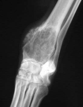
Osteosarcoma refers to the most common bone tumor found in dogs. Bone cancer can affect any breed of dog, but it is more commonly found in the larger breeds (ie., American Bulldog, Golden Retriever, Labrador Retriever, Rottweiler, Great Dane)

The disease is extremely aggressive and has a tendency to spread rapidly into other parts of the dog's body (metastasize). There are treatment options available, but generally the long term prognosis for the animal is poor.

Bone cancer can be found in cats as well, but it is rare.  

Symptoms and Types
Many signs of bone cancer are subtle. They can include swelling, lameness, and joint or bone pain. In some cases, dogs suffering from bone cancer will appear tired or have anorexia. Occasionally, dogs will exhibit a mass growth on their body or a painful inflammation around the sight of the tumor.

Causes
Current knowledge of the disease has not linked genetics or gender to the condition, but bone cancer does appear more often in large to giant breeds of dogs. Some studies have shown a slight increase in the development of osteosarcoma among dogs that have experienced a blunt bone injury.

Diagnosis
Your veterinarian will use X-rays to view the mass, often using several angles to get an accurate picture. Other tests include biopsies, blood tests, bone scans, and CAT scans to view the bone areas, and the mass, if discovered. If the diagnosis is bone cancer, it is important to note that the prognosis is often unfavorable and that there are numerous side effects to the treatment options. Management on your part will be required.

Treatment
Chemotherapy is often used as a supplement to any surgical options to ensure that the disease has not spread into other areas of the dog's body, particularly the lymph nodes. In severe cases, limbs may need to be amputated to completely remove the bone cancer.

Living and Management
Activity will be restricted following any surgery. A pain management program and medications are frequently prescribed for the animal following surgery. Medications commonly work to manage pain and reduce inflammation. Ongoing management and monitoring of the dog's white and red blood cell counts will be recommended, and chest X-rays are often used to determine remission.

Prevention
There are currently no known prevention methods for bone cancer.


0 Comments

Eyelash Disorders in Dogs

10/30/2014

0 Comments

 
Picture
Picture
Picture
Trichiasis, Distichiasis, and Ectopic Cilia in Dogs  

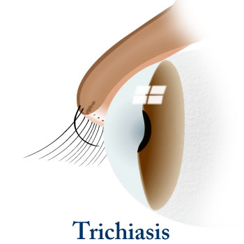
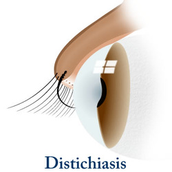
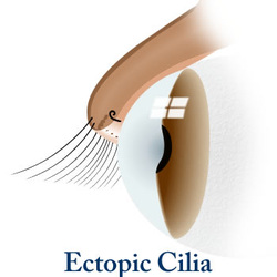
Trichiasis, distichiasis, and ectopic cilia are eyelash disorders that are found in dogs. Trichiasis is in-growth of the eyelashes; distichiasis is an eyelash that grows from an abnormal spot on the eyelid; and ectopic cilia are single or multiple hairs that grow through the inside of the eyelid. In all of these conditions, the eyelash hair can come into contact with and damage the cornea or conjunctiva of the eye.

These conditions are commonly seen in young dogs but dogs of any age or breed may be affected. However, trichiasis does tend to appear more frequently in Pekingese, English cocker spaniel, pugs, and bulldogs; distichiasis is common in cocker spaniels, miniature long haired dachshunds, English bulldogs, golden retrievers, toy and miniature poodles, Shetland sheepdogs and Pekingese; and ectopic cilia are more commonly found in dachshunds, lhasa apsos, shih tzus, boxers, golden retrievers, and Shetland sheepdogs.

Symptoms and Types Trichiasis
  • Change in pigmentation of the iris (colored portion of the eye)
  • Abnormal ticking or twitching of the eyelid (blepharospasm)
  • Overflow of tears
  • Swelling of eyes

Distichiasis
  • Mostly no symptoms can be seen
  • Stiff cilia (eyelash)
  • Pawing at eye
  • Abnormal tick or twitch of eyelid (blepharospasm)
  • Overflow of tears (epiphora)
  • Increased blood vessels in the cornea
  • Change in iris pigmentation
  • Corneal ulcers

Ectopic cilia
  • Eye pain
  • Severe abnormal ticking or twitching of the eyelid (blepharospasm)
  • Overflow of tears (epiphora)

Causes
  • Facial conformation and breed predisposition
  • Unknown etiology in many cats

Diagnosis
Your veterinarian will carefully inspect the eye structures and eyelashes to discern exactly which eyelash disorder your dog has. The diagnosis is usually straightforward in most cases. Your veterinarian will perform a Schirmer tear test to measure tear production and evaluate whether the affected eye is producing enough tears to keep it moist, and a fluorescein stain over the surface of the eye to make corneal ulcers visible. Determination of intraocular (within the eye) pressure is also an important test in evaluating the eye. This test will allow your veterinarian to evaluate the level of fluid pressure inside the eye. More specific testing may be performed to evaluate both the superficial and deep structures of eye.

Treatment
Eliminating the eye irritation will resolve symptoms in most cases. In case of trichiasis, in some patients the hair will be cut short to prevent eye irritation. In others patients surgery may be required for correction of the defect.

In distichiasis no treatment is usually required. Hairs that have been mechanically plucked will regrow within four to five weeks, and will need to removed again. In some cases of distichiasis, surgery may be required. For example, in cases where the hair is a frequent irritation to the surface of the eye.

In case of ectopic cilia, surgery is the preferred method for removing the ectopic eyelash hairs.

Living and Management

Observe your dog's eyes and consult your veterinarian if you notice any recurrence of symptoms. Keep the eyes clean, either with fresh water, or with a veterinarian recommended eye wash. In cases of distichiasis, re-growth is common, in which case you may need to revisit your veterinarian for follow-up care.

0 Comments

Pyometra and Cystic Endometrial Hyperplasia in Dogs

9/4/2014

0 Comments

 
Picture
Picture
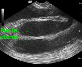
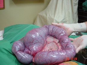
The abnormal thickening (pyometra) of the uters' lining can occur in dogs at any age, although it is more common in dogs that are six years of age or older. Cystic endometrial hyperplasia, meanwhile, is a medical condition characterized by the presence of pus-filled cyst inside the dog's uterus, causing the endometrium to enlarge (also known as hyperplasia).

Prognosis is often positive for both conditions; however, if the dog's cervix is closed, it can be a life threatening condition requiring immediate medical attention.

Symptoms and Types
Signs may include:
  • Abdominal distention (from an enlarged uterus)
  • Vulvar (vaginal) discharge
  • Closed cervix
  • Lethargy
  • Depression
  • Lack of appetite
  • Vomiting
  • Frequent urination

Causes

One of the known causes of this condition in dogs is repeat exposure to estrogen andprogesterone. The formation of cystic endometrial hyperplasis is often progressive, often following the development of a thickened uterine lining.

Intact older female dogs that have never given birth are at a higher risk of developing pyometra or cystic endometrial hyperplasia.

Diagnosis
Your veterinarian will perform an examination to review the type and severity of your dog’s discharge, as well as to view whether the cervix is open or closed. X-rays and ultrasounds will be used detect the size of the uterus, and to determine if the dog is pregnant.

Treatment
In many cases, treatment for pyometra will be given on an outpatient basis. However, if the cervix is closed, the condition can be life threatening and immediate action will be required. The preferred treatment for this medical condition is a hysterectomy -- the removal of the dog's ovaries and uterus. Other options are available, but at a higher risk to the animal's wellbeing; these are only recommended for dogs with a high breeding value.

A lavage of the uterus and surrounding areas will be performed to remove the pus and fluids, and to support the healing process. Antibiotics are often administered to fight off infection. Prostaglandins, meanwhile, are administered to control the dog's cell growth and control hormone regulation, and to cause the smooth muscles in the dog's body to contract.

Living and Management
Your dog will be released from medical care once its uterus has returned to normal size and there are no signs of fluids. Antibiotics should be administered for several weeks to prevent infection. It is normal for vaginal discharge to continue until the healing process is complete.

Prevention
Allowing your dog to go through its heat (estrus) cycles without being bred has been shown to increase the incidence of pyometra. Therefore, spaying your dog (or removing its ovaries) is the best form of prevention.

0 Comments

Seizures (Epileptic) in Dogs

8/20/2014

2 Comments

 
Epilepsy, Idiopathic or Genetic, in DogsEpilepsy is a brain disorder that causes the dog to have sudden, uncontrolled, recurring physical attacks, with or without loss of consciousness. This may sometimes occur for unknown reasons (idiopathic) or due to genetic abnormalities. However, idiopathic epilepsy is often characterized by structural brain lesions and is more likely seen in male dogs. If left untreated, the seizures may become more severe and frequent.

Symptoms and Types
Seizures are usually preceded by a short aura (or focal onset). When this occurs the dog may appear fightened and dazed, or it may hide or seek attention. One theseizure(s) begin, the dog will fall on its side, become stiff, chomp its jaw, salivate profusely, urinate, defecate, vocalize, and/or paddle with all four limbs. These seizure activities generally last between 30 and 90 seconds.

Seizures most often occur while the patient is resting or asleep, often at night or in early morning. In addition, most dogs recover by the time you bring the dog to the veterinarian for examination.

Generally, the younger the dog is, the more severe the epilepsy will be. As a rule, when onset is before age 2, the condition responds positively to medication. Behavior following the seizure, known as postictal behavior, include periods of confusion and disorientation, aimless wandering, compulsive behavior, blindness, pacing, increased thirst (polydipsia) and increased appetite (polyphagia). Recovery following the seizure may be immediate, or it may take up to 24 hours.

Dogs with established epilepsy can have cluster seizures at regular intervals of one to four weeks. This is particularly evident in large-breed dogs.

Causes
Idiopathic epilepsy is genetic in many dog breeds and is familial; meaning that it runs in certain families or lines of animals. Breeds most prone to idiopathic epilepsy include the Beagle, Dachshund, Keeshond, Belgian Tervuren, Golden Retriever, Labrador Retriever, Vizsla and Shetland Sheepdog. Multiple genes and recessive modes of inheritanceare suggested in the Bernese Mountain Dog and Labrador Retriever, while non-gender hormone recessive traits has been proposed in the Vizsla and Irish Wolfhound. There are also recessive traits in the English Springer Spaniel which can lead to epilepsy, but it does not appear to affect all members of the family. Seizures are mainly focal (involving localized areas of the brain) in the Finnish Spitz.

The characteristics associated with genetic epilepsy usually manifests from 10 months to 3 years of age, but has been reported as early as six months and as late as five years.

Diagnosis
The two most important factors in the diagnosis of idiopathic epilepsy is the age at onset and the seizure pattern (type and frequency). If your dog has more than two seizures within the first week of onset, your veterinarian will probably consider a diagnosis other than idiopathic epilepsy. If the seizures occur when the dog is younger than six months or older than five years, it may be metabolic or intracrainal (within the skull) in origin; this will rule out hypoglycemia in older dogs. Focal seizures or the presence of neurologic deficits, meanwhile, indicate structural intracranial disease.

Treatment
Most of the treatment is outpatient. It is recommended that the dog does not attempt to swim, to prevent accidental drowning white it undergoes treatment. Be aware that most dogs on long-term antiepileptic or anticonvulsant medications become overweight. Therefore, monitor its weight closely and consult your veterinarian for a diet plan if necessary.

Living and Management
It is essential to monitor therapeutic levels of drugs in the blood. Dogs treated with phenobarbital, for instance, must have their blood and serum chemistry profile monitored after initiating therapy during the second and forth week. These drug levels will then be evaluated every 6 to 12 months, changing the serum levels accordingly.

Carefully monitor older dogs with kidney insufficiency that are on potassium bromide treatment; your veterinarian may recommend a diet change for these dogs.

Prevention
Because this form of epilepsy is due to genetic abnormalities, there is little you can due to prevent them. However, the abrupt discontinuation of medication(s) to control seizures may aggravate or initiate seizures. Additionally, avoid salty treats for dogs treated with potassium bromide, as it may lead to seizures.

2 Comments

Blindness in Dogs - "Quiet Eye" (No redness)

8/4/2014

1 Comment

 
Blind quiet eye is the loss of vision in one or both eyes without ocular vascular injection or other apparent signs of eye inflammation. This may occur due to abnormalities in retinal image detection, retinal focusing, optic nerve transmission, or simply the central nervous system's inability to interpret images correctly.

Symptoms and Types
Because Blind Quiet Eye directly affects the dog's vision, it may display several signs, including:

  • Clumsy behavior (e.g., bumping into objects, tripping, falling)
  • Decreased or absent menace response (i.e., does not blink when a hand is waved toward the eyes)
  • Impaired visual placing responses (e.g., extends the paws incorrectly when trying to approach a nearby surface)

In addition, these problems may become more exaggerated when the dog is outside at night.

Causes
There are several causes for Blind Quiet Eye, such as cataracts, central nervous system lesions, and the lens' inability to focus correctly. Other common causes include:

  • Retinal disorders:
    • Sudden acquired retinal degeneration syndrome (SARDS) - common breeds affected include American cocker spaniel, Bichon frise, Brittany spaniel, Dachshund, English springer spaniel, Miniature schnauzer, Pug
    • Shrinking of the retina (progressive retinal atrophy) - common breeds affected include Miniature schnauzer, Maltese, Old English sheepdog, Cardigan Welsh corgi, Border collie, American pit bull terrier, Poodle, Great Dane, Bernese Mountain dog, Rottweiler, Labrador retriever, Shih tzu, Irish setter, Mastiff, Siberian husky, Yorkshire terrier, Australian shepherd, German shepherd dog, Golden retriever, Portuguese water dog 
    • Separation of the eye's inner lining (retinal detachment) - common breeds affected include Labrador retriever, Poodle, Shih tzu, Collie
    • Ivermectin toxicity - common breeds affected include Border collie, Old English sheepdog, Collie, Shetland sheepdog
  • Optic nerve issues due to:
    • Inflammation
    • Cancer
    • Trauma
    • Underdevelopment
    • Lead Toxicity

Diagnosis
You will need to give a thorough history of your dog’s health and the onset and nature of the symptoms to the veterinarian. He or she will then perform a complete physical examination (including an opthalmoscopic exam) as well as a biochemistry profile,urinalysis, complete blood count (CBC) to rule out potential systemic causes of the disease.

During the ophthalmic exam a penlight will be used to rule out potential systemic causes of the disease, such as cataracts or retinal detachment. (In cases of retinal detachment, the systemic blood pressure is often elevated.) Ophthalmoscopy, meanwhile, may reveal progressive retinal atrophy or optic nerve disease.

If the ophthalmic exam reveals nothing irregular, it may suggest sudden acquired retinal degeneration syndrome (SARDS), retrobulbar optic neuritis (inflammation of the optic nerve after it exits the eye toward the brain), or a central nervous system (CNS) lesion. If the diagnosis is still in doubt, electroretinography -- whic measures the electrical responses of photoreceptor cells in the retina -- makes it possible to differentiate retinal from optic nerve or CNS disease. Ocular ultrasounds and CT (computed tomography) and MRI (magnetic resonance imaging) scans are also very helpful to visualize and diagnose orbital or CNS lesions.

Treatment
Your veterinarian will try to localize the disease and will often refer you to a veterinary ophthalmologist. Unfortunately, there is no effective treatment for Blind Quiet Eye brought on by SARDS, progressive retinal atrophy, optic nerve atrophy, or optic nerve hypoplasia. However, cataracts, luxated lenses, and some forms of retinal detachment may be treated surgically.

In addition, dogs with retinal detachment should have their exercise severely restricted until the retina is firmly reattached. These patients should also be switched to a calorie-restricted diet to prevent obesity, which could occur due to reduced activity.

Living and Management
With assistance, blind pets can lead relatively normal and functional lives. However, dogs with progressive retinal atrophy or genetic cataracts should not be bred. Your veterinarian will recommend you with some basic safety concepts, such as examining for potential hazards in your home. He or she will also schedule regular follow-up exams to ensure that any ocular inflammation is controlled and to ensure, if possible, that your pet’s vision is maintained.

1 Comment

Patellar Luxation - Kneecap Dislocation in Dogs

7/22/2014

1 Comment

 
Picture
Patellar Luxation in Dogs
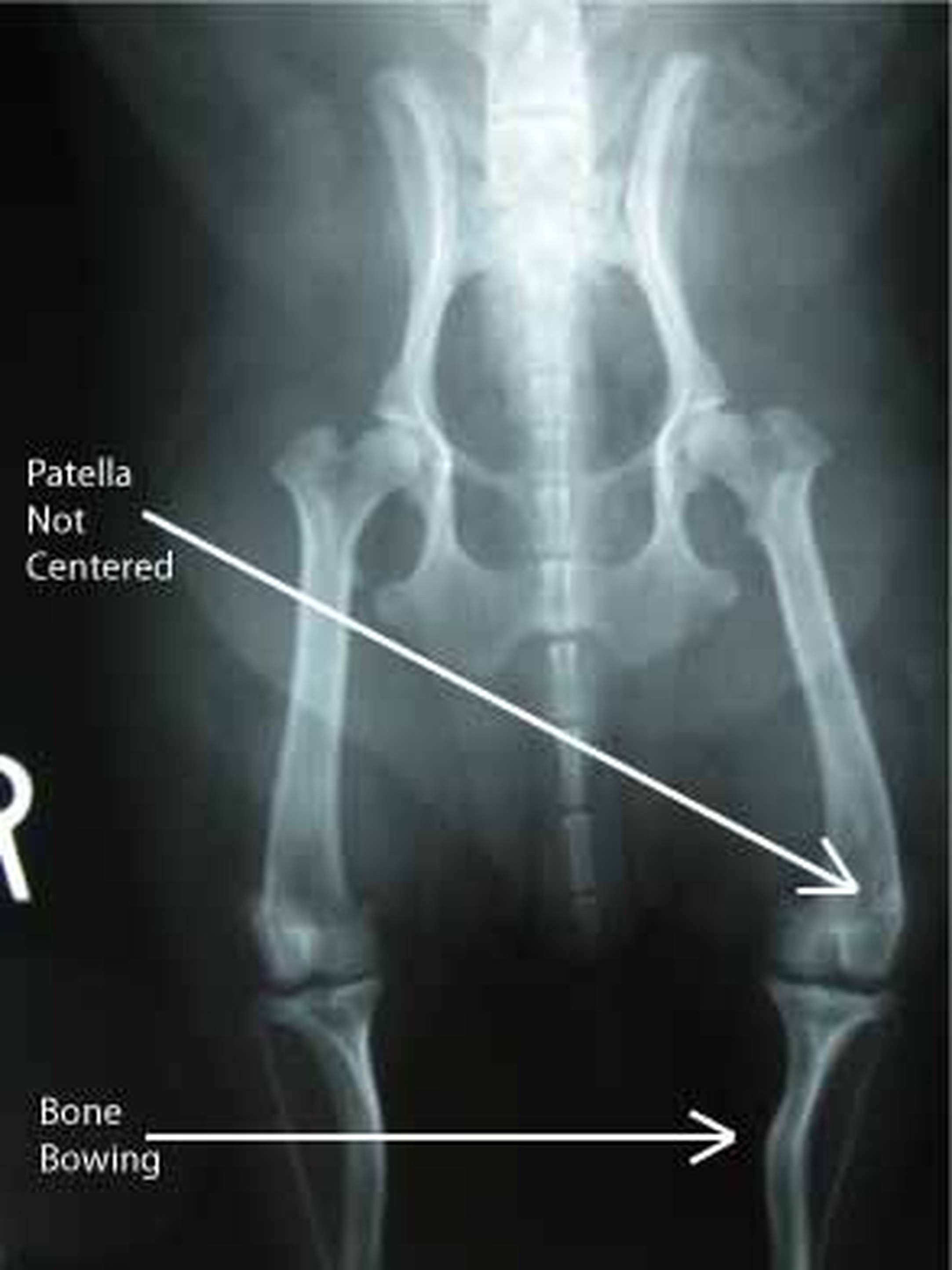
Patellar luxation occurs when the dog's kneecap (patella) is dislocated from its normalanatomic position in the groove of the thigh bone (femur). When the kneecap is dislocated from the groove of the thigh bone, it can only be returned to its normal position once the quadriceps muscles in the hind legs of the animal relax and lengthen. It is for this reason that most dogs with the condition will hold up their hind legs for a few minutes.

A dislocated kneecap is one of the most prevalent knee joint abnormalities in dogs. The condition is most common in toy and miniature dog breeds such as the Yorkshire Terrier, Dachshund, West Highland White Terrier (Westie), Pomeranian, Pekingese, Chihuahua, and Boston Terrier. Female dogs are 1 1/2 times more likely to acquire the condition.

Symptoms and Types
The specific symptoms of a dislocated kneecap will depend on the severity and persistence of the condition, as well as the amount of degenerative arthritis that is involved. Typically, a dog with a dislocated kneecap will exhibit prolonged abnormal hindlimb movement, occasional skipping or hindlimb lameness, and sudden lameness.

The dog will rarely feel pain or discomfort once the kneecap is out of position, only feeling pain at the moment the kneecap slides out of the thigh bone's ridges.

Causes
A dislocated kneecap is usually caused by a genetic malformation or trauma. The clinical signs of the condition will normally start showing approximately four months after birth.

Diagnosis
A dislocated kneecap is diagnosed through a variety of means. Top view (craniocaudal) and side view (mediolateral) X-rays of the stifle joint, hip, and hock may be used to detect bending and twisting of the thigh bone and larger bone of the lower leg. Skyline X-rays may reveal a shallow, flattened, or curved groove of the thigh bone. A fluid sample taken from the joint and an analysis of the lubricating fluid in the joint (synovial fluid) will show a small increase in mononuclear cells. It is also necessary for the veterinarian to perform an examination by touch to feel for kneecap freedom.

Treatment
Medical treatment for kneecap dislocation has very little effectiveness; surgery is the preferred treatment of choice for severe cases. Surgery can correct both the affected structures and the movement of the kneecap itself, and in 90 percent of cases, frees the dog from lameness and dysfunction.

The kneecap may be fastened on the outside of the bone to prevent it from sliding towards the inside. Alternatively, the groove of the thigh bone may be deepened so that it can better hold the kneecap.

Living and Management
Follow-up treatment after successful surgery will include leash walk exercise for one month (avoid jumping) and yearly examinations to check for progress. It is important that pet owners are aware that there is a high possibility of recurrence (48 percent), although the dislocation will be considerably less severe than the original incidence. Because kneecap dislocation is genetically inherited, the breeding of affected dogs is highly discouraged.

PreventionThere are currently no known preventative measures for this medical condition.

1 Comment
<<Previous
    PET SCAN Blog: 
    The INNER Pet... 
    Pet Health Inside & Out
    The goal of this blog is to help educate pet owners by sharing pet health facts and interesting pet news articles

    Archives

    September 2019
    August 2019
    June 2016
    March 2016
    November 2015
    June 2015
    April 2015
    March 2015
    January 2015
    December 2014
    November 2014
    October 2014
    September 2014
    August 2014
    July 2014
    June 2014
    May 2014
    April 2014
    March 2014
    February 2014
    January 2014

    Categories

    All
    Cancer In Pets
    Cat Health
    Dog Health
    Fun Facts
    Pets In The News
    Research News
    Videos

    RSS Feed

"YOUR Partner for Advanced Diagnostic Excellence"
Providing mobile ultrasound consultation services to veterinarians in Chandler, Gilbert, Mesa, Tempe, Ahwatukee, Scottsdale, and the Greater Phoenix Metropolitan Area. 
Picture


Proud member of the
American Veterinary Medical Association