Case Spotlight: Heartworm Disease
|
History: An intact male mixed breed dog was presented for evaluation of 4-5 days of progressive abdominal distension. Abnormalities on laboratory work were heartworm positive and anemia. Survey radiographs showed cardiomegaly and a possible abdominal mass.
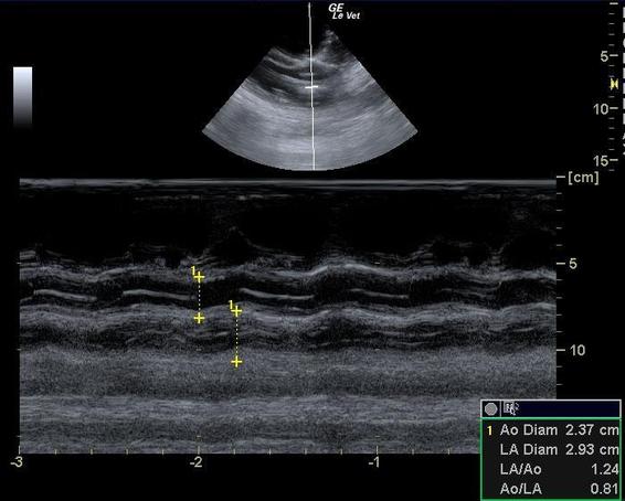
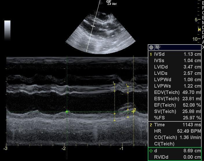
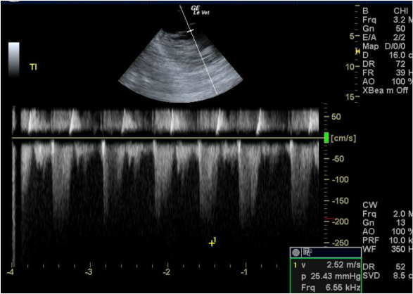
Clinical Differential Diagnosis: Cardiac - right heart failure secondary to heartworm, pericardial effusion, cardiomyopathy Abdominal mass - neoplasia/granuloma/abscess/cyst of spleen, liver, kidney. Sonographic Interpretation: The echocardiogram revealed an enlarged right heart with right atrial enlargement and significant tricuspid regurgitation. Tricuspid insufficiency was noted at 3.87 m/sec. The main branch of the pulmonary artery was enlarged and a large amount of heartworms were noted. Smoke was noted in the right atrium. The mitral valve was insufficient. The left atrium was of normal size. Tricuspid insufficiency velocity 3.87 m/sec. Mitral insufficiency velocity 5.65 m/sec. Sonographic Differential Diagnosis: Right sided heart failure. Large amount of heartworms noted in the main branch of the pulmonary artery. Pulmonary hypertension was noted with a jet of 3.87 m/sec. Given the changes seen on echocardiographic examination this patient would be an ideal candidate for basket retrieval of heartworms. Until this procedure is performed the patient should be treated with aspirin at 1 mg/kg every other day, Pimobendan at 0.25-0.3 mg/kg b.i.d. and Sildenafil at 1 mg/kg b.i.d. The Sildenafil dose can be increased to 1.5 mg/kg b.i.d. over 10 days. Lasix should be instituted at 1-2 mg s.i.d. Given the significant amount of heartworms noted in the pulmonary artery coupled with the patient's clinical signs the patient's prognosis is guarded at this time. Ideally, it would be prudent to perform a blood pressure and ECG in this patient. Diagnosis: Heartworms, pulmonary hypertension.
|
Patient Information
Age 5 Year Gender Male, Intact Species Canine Clinical Signs
Blood Chemistry
Special Testing
Call (480) 760-5878 to schedule a mobile
ultrasound consultation c SAME-DAY & STAT Appointments Available |