Pet Scan:  Mobile Veterinary Ultrasound Service
  • Home
  • Indications for Ultrasound
  • Services
  • What To Expect
  • Our Doctors
  • Referring DVMs

Using Viruses to Treat Cancer in Pets

12/12/2014

1 Comment

 
Surgery, radiation, and chemotherapy are the more commonly known treatments for cancer in pets. But newer technologies are opening up other possibilities. A recent experiment summary (abstract) suggests the use of genetic modified viruses to treat various types of cancer.

Oncolytic Virotherapy
The idea of using a virus for cancer treatment or oncolytic virotherapy is not a new idea. In the 1940s scientists conducted animal studies using viruses to treat tumors. Doctors in the 1950s observed that cancer patients that were stricken with viral infections or recently vaccinated experienced improvement in their condition. It was believed that the infections or vaccinations triggered an immune response that increased the production of interferon and tumor necrosis factors, or TNFs.

Interferons are large molecules released by cells infected with viruses, bacteria, parasites and tumors to interfere, hence their name, with virus reproduction and to trigger responses from immune cells. Interferons activate natural killer white blood cells and large white cells called macrophages that attack and destroy the invading organisms and cancer cells. Interferon promotes the production or molecular complexes that attach to viral, bacterial, parasite, and tumor cells so they are more quickly and effectively attacked by killer white cells. TNFs causes destructive changes in cell walls and causes foreign or tumor cells to burst and die

Despite the potential for viral therapy of cancer in these early years, it required the present advances in technology to achieve a real possibility. Precisely, it required our present ability to genetically modify organisms like viruses and safely use them to target cancer cells. The viruses are modified to prevent their normal ability to cause disease and genetically altered to produce interferon or other anti-cancer molecules.

Preliminary Study in Dogs
The abstract was based on a small study intended to evaluate the safety and effectiveness of a new oncolytic virus. The group was made up of of seven dogs suffering from various cancers (lymphoma, malignant melanoma and multiple myeloma). The researchers used a novel virus for their study; they used a modified vesicular stomatitis virus that causes oral, udder, and hoof ulcers in cattle. Although seldom fatal, the disease causes inappetence and decreased milk or meat production [in cattle]. It can also infect horses and pigs, and rarely, sheep, goats, and llamas. Because of its effect on agricultural production, vesicular stomatitis is a diagnosis that requires mandatory reporting to federal and state animal health officials.

The virus was also modified to produce human or canine interferon. Three dogs received the human form and four dogs received the canine form. The abstract reported measurable improvement but did not specify the type and extent of the improvements except for the production of neutralizing antibodies within 7-10 days after viral administration. Side effects were minimal and included reversible changes in liver enzymes, fever, and urinary tract infection. Virus was not shed in the urine or saliva. These limited side effects are comparable or even fewer than those expected with radiation or chemotherapy.

This is a small study and rightly titled as preliminary. It has yet to be published so critical evaluation is still not available. Clearly, much more study is required for this type of treatment. What is exciting is that this is one of many new potential treatments for cancer in pets. Advanced cancer treatment in the last decade has changed how the diagnosis is now viewed. Rather than an immediate death sentence, cancer can now be better managed as a chronic disease much like kidney and heart conditions. These new treatments offer greater treatment flexibility and potentially an improved quality of life.

1 Comment

Bone Cancer (Osteosarcoma) in Dogs

11/14/2014

0 Comments

 
Picture
Osteosarcoma in Dogs
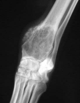
Osteosarcoma refers to the most common bone tumor found in dogs. Bone cancer can affect any breed of dog, but it is more commonly found in the larger breeds (ie., American Bulldog, Golden Retriever, Labrador Retriever, Rottweiler, Great Dane)

The disease is extremely aggressive and has a tendency to spread rapidly into other parts of the dog's body (metastasize). There are treatment options available, but generally the long term prognosis for the animal is poor.

Bone cancer can be found in cats as well, but it is rare.  

Symptoms and Types
Many signs of bone cancer are subtle. They can include swelling, lameness, and joint or bone pain. In some cases, dogs suffering from bone cancer will appear tired or have anorexia. Occasionally, dogs will exhibit a mass growth on their body or a painful inflammation around the sight of the tumor.

Causes
Current knowledge of the disease has not linked genetics or gender to the condition, but bone cancer does appear more often in large to giant breeds of dogs. Some studies have shown a slight increase in the development of osteosarcoma among dogs that have experienced a blunt bone injury.

Diagnosis
Your veterinarian will use X-rays to view the mass, often using several angles to get an accurate picture. Other tests include biopsies, blood tests, bone scans, and CAT scans to view the bone areas, and the mass, if discovered. If the diagnosis is bone cancer, it is important to note that the prognosis is often unfavorable and that there are numerous side effects to the treatment options. Management on your part will be required.

Treatment
Chemotherapy is often used as a supplement to any surgical options to ensure that the disease has not spread into other areas of the dog's body, particularly the lymph nodes. In severe cases, limbs may need to be amputated to completely remove the bone cancer.

Living and Management
Activity will be restricted following any surgery. A pain management program and medications are frequently prescribed for the animal following surgery. Medications commonly work to manage pain and reduce inflammation. Ongoing management and monitoring of the dog's white and red blood cell counts will be recommended, and chest X-rays are often used to determine remission.

Prevention
There are currently no known prevention methods for bone cancer.


0 Comments

Eyelash Disorders in Dogs

10/30/2014

0 Comments

 
Picture
Picture
Picture
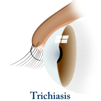
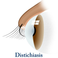
Trichiasis, Distichiasis, and Ectopic Cilia in Dogs  

Trichiasis, distichiasis, and ectopic cilia are eyelash disorders that are found in dogs. Trichiasis is in-growth of the eyelashes; distichiasis is an eyelash that grows from an abnormal spot on the eyelid; and ectopic cilia are single or multiple hairs that grow through the inside of the eyelid. In all of these conditions, the eyelash hair can come into contact with and damage the cornea or conjunctiva of the eye.

These conditions are commonly seen in young dogs but dogs of any age or breed may be affected. However, trichiasis does tend to appear more frequently in Pekingese, English cocker spaniel, pugs, and bulldogs; distichiasis is common in cocker spaniels, miniature long haired dachshunds, English bulldogs, golden retrievers, toy and miniature poodles, Shetland sheepdogs and Pekingese; and ectopic cilia are more commonly found in dachshunds, lhasa apsos, shih tzus, boxers, golden retrievers, and Shetland sheepdogs.

Symptoms and Types Trichiasis
  • Change in pigmentation of the iris (colored portion of the eye)
  • Abnormal ticking or twitching of the eyelid (blepharospasm)
  • Overflow of tears
  • Swelling of eyes

Distichiasis
  • Mostly no symptoms can be seen
  • Stiff cilia (eyelash)
  • Pawing at eye
  • Abnormal tick or twitch of eyelid (blepharospasm)
  • Overflow of tears (epiphora)
  • Increased blood vessels in the cornea
  • Change in iris pigmentation
  • Corneal ulcers

Ectopic cilia
  • Eye pain
  • Severe abnormal ticking or twitching of the eyelid (blepharospasm)
  • Overflow of tears (epiphora)

Causes
  • Facial conformation and breed predisposition
  • Unknown etiology in many cats

Diagnosis
Your veterinarian will carefully inspect the eye structures and eyelashes to discern exactly which eyelash disorder your dog has. The diagnosis is usually straightforward in most cases. Your veterinarian will perform a Schirmer tear test to measure tear production and evaluate whether the affected eye is producing enough tears to keep it moist, and a fluorescein stain over the surface of the eye to make corneal ulcers visible. Determination of intraocular (within the eye) pressure is also an important test in evaluating the eye. This test will allow your veterinarian to evaluate the level of fluid pressure inside the eye. More specific testing may be performed to evaluate both the superficial and deep structures of eye.

Treatment
Eliminating the eye irritation will resolve symptoms in most cases. In case of trichiasis, in some patients the hair will be cut short to prevent eye irritation. In others patients surgery may be required for correction of the defect.

In distichiasis no treatment is usually required. Hairs that have been mechanically plucked will regrow within four to five weeks, and will need to removed again. In some cases of distichiasis, surgery may be required. For example, in cases where the hair is a frequent irritation to the surface of the eye.

In case of ectopic cilia, surgery is the preferred method for removing the ectopic eyelash hairs.

Living and Management

Observe your dog's eyes and consult your veterinarian if you notice any recurrence of symptoms. Keep the eyes clean, either with fresh water, or with a veterinarian recommended eye wash. In cases of distichiasis, re-growth is common, in which case you may need to revisit your veterinarian for follow-up care.

0 Comments

Bladder Inflammation in Cats

10/8/2014

0 Comments

 
Picture


Interstitial Cystitis in Cats, Feline Interstitial Cystitis (FIC)

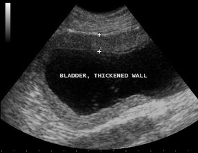
Feline interstitial cystitis, sometimes called feline idiopathic cystitis or FIC, is an inflammation of the bladder that causes symptoms of lower urinary tract disease. However, in the case of interstitial cystitis, a definitive cause for the disease cannot be identified.

Feline interstitial cystitis can occur in both female and male cats. It is a chronic disease that can be difficult to treat and frustrating for cats and cat owners alike.

Symptoms and Types
Symptoms associated with interstitial cystitis include:

  • Frequent attempts to urinate
  • Straining to urinate
  • Urinating in inappropriate places in the house
  • Crying out during attempts to urinate
  • Blood-tinged urine

Causes

Though the cause of feline interstitial cystitis is not fully understood, stress and the changes inherent in the body as a result of stress are thought to play a large part in interstitial cystitis. Some researchers believe that interstitial cystitis is only one of the manifestations seen in cats suffering from stress and may only be the “tip of iceberg” in terms of symptoms that may be caused by stress. Abnormalities have also been found in the nervous, endocrine and cardiovascular systems of cats in addition to the urinary system. It remains unknown why some cats develop symptoms of FIC and others do not.

Diagnosis
Diagnosis relies on ruling out other diseases that may cause similar symptoms, such as urinary tract infections, bladder stones, and other bladder abnormalities in cats. Testing that is frequently performed include:

  • A blood screen, including a complete blood cell count (which examines the different types of cells circulating in the blood stream, such as red blood cells and white blood cells) and chemistry profile (which is useful in evaluating the function of major organs such as the liver and kidneys)
  • A urinalysis, which checks for abnormalities in the urine, including blood, crystals, protein and other abnormal substances as well as testing the pH (which determines how acidic the urine is),  and the urine specific gravity (which determines whether the urine is concentrated or not)
  • An abdominal X-ray and/or an ultrasound exam of the bladder to rule out stones and other abnormal structures in the bladder
TreatmentTreatment consists primarily of modifying the environment to help reduce stress levels, dietary modifications, medications for pain, and other pharmaceuticals that may alter your cat’s mental state.

Multi-modal environmental modifications (MEMO) is the term that is used to describe altering the cat’s environment in an attempt to reduce the cat’s stress level. See the Living and Management section below for more information about MEMO.

Pain medications are often used in treating interstitial cystitis to relieve the discomfort caused by the inflammation within the bladder and urinary tract and make your cat more comfortable while urinating.

Dietary modifications are often necessary and you should follow your veterinarian’s directions if a special diet is recommended. Increasing water consumption is an important part of treatment and feeding canned food, when possible, will help increase the moisture content in your cat’s diet.

Pheromones such as Feliway are often recommended to help reduce stress levels for your cat.

Other medications that your veterinarian may advise include amitriptyline, clomipramine or fluoxetine, all of which are antidepressants. These medications are generally reserved for cases where MEMO, dietary modifications and pain medications have failed to help.

Living and Management
MEMO involves providing for all of your cat’s basic needs.

Litter box management is a necessary part of environmental modification.

  • Provide an adequate number of litter boxes. There should be one more litter box than the number of cats in the household.
  • Most cats prefer a large litter box over a smaller one.
  • Make sure the sides of the litter boxes are not too high for your cat. This is especially important for older cats that may suffer from arthritis or other mobility issues and for young kittens.
  • Choose an appropriate litter for your cat. The ideal litter is dust-free and fragrance-free. Strongly scented litters may be pleasant for you but not for your cat. You may have experiment with different types of litters to find the one your cat prefers.
  • Be sure to clean the litter boxes frequently.
  • Place all litter boxes in a quiet location where your cat will not be disturbed or frightened when using the box.
Provide feeding and water stations that are accessible for all cats in your household. If you have more than one cat, you may need to provide more than one feeding and water station.

Cats prefer perches, preferably at eye-level or above, on which to rest and observe their surroundings. Be sure to provide an adequate number of perches for all the cats in your household. Consider placing one or more of these perches near a window for your cat’s enjoyment.

All cats need hiding places. Your cat should have a place to retreat where he will not be disturbed by people or by other pets. In a multi-cat household, be sure there is an adequate number of hiding places for all cats.

Provide interactive toys for your cat. Food puzzles are also helpful to provide distraction for your cat and also provide exercise.


0 Comments

Aspirin Poisoning in Cats

10/8/2014

0 Comments

 
Aspirin Toxicity in Cats
Aspirin, a non-steroidal anti-inflammatory medication, has been found to have beneficial effects for some animals. It has been used for conditions related to blood clotting, inflammation, and for its analgesic properties. However, it can also be toxic to the body. Once ingested, aspirin forms salicylic acid, which is then distributed throughout the body. Aspirin toxicity is a particular concern in cats because they lack the enzyme critical for metabolizing salicylic acid properly. Cat owners must follow their veterinarian's orders strictly if aspirin is prescribed for any reason.

Symptoms and Types
The progression of symptoms can occur quickly. One of the first noticeable signs is loss of appetite. Other signs include vomiting, diarrhea, and intestinal hemorrhagebrought on by ulceration in the stomach and small intestines. The central nervous system may also be affected, causing your cat to have trouble walking, appear weak and uncoordinated, or even collapse. Loss of consciousness and sudden death can also occur.

Since even nontoxic levels can produce these symptoms, You will need to monitor your cat for any digestive problems or changes in behavior when giving your cat aspirin for any medical reason. If a significant amount of aspirin is ingested, emergency medical treatment will be necessary.

Diagnosis
If you know, or even suspect that your cat has ingested aspirin, and your cat is showing apparent symptoms of toxicity, diagnostic tests should focus on the severity of the toxicity. A complete blood profile will be conducted, including a chemical blood profile, a complete blood count, and a urinalysis. Usually an affected cat will be anemic, with electrolyte abnormalities, in addition to showing a reduction in the blood's ability to clot properly.

Treatment
Cats treated within 12 hours of ingestion, and which are presenting limited signs of distress, can have the concentration of aspirin in the body decreased through a prescribed treatment of decontamination. The sooner this care begins, the better. Your veterinarian may recommend that you decrease the amount of aspirin in the body by inducing vomiting at home before coming to the clinic, or the vomiting may be induce in the clinic. By inducing vomiting, or pumping the stomach (gastric lavage), your veterinarian will be able to remove as much aspirin as possible, lowering the chances of permanent injury. Activated charcoal may be given after vomiting to absorb some of the remaining aspirin.

Medications to encourage healing, or to protect the gastrointestinal lining are also generally prescribed. Depending on your cat's status, fluids and other supportive treatments may also be necessary. Hospitalization and repeated blood analysis will often be standard until your cat is stable.

Living and Management
Aspirin has several clinical uses. It can be prescribed as a pain reliever, an anti-inflammatory, an anti-platelet blood thinning agent, and for lowering an abnormal body temperature. If aspirin is being used for a chronic condition, such as for preventing a blockage of the blood vessel (arterial thromboembolism), it is important to follow your veterinarian’s directions. Reducing or discontinuing the aspirin dosage may be necessary if your pet is showing a susceptibility to toxicity.

0 Comments

Pyometra and Cystic Endometrial Hyperplasia in Dogs

9/4/2014

0 Comments

 
Picture
Picture
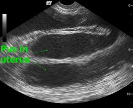
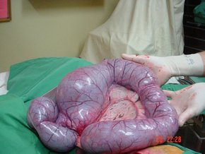
The abnormal thickening (pyometra) of the uters' lining can occur in dogs at any age, although it is more common in dogs that are six years of age or older. Cystic endometrial hyperplasia, meanwhile, is a medical condition characterized by the presence of pus-filled cyst inside the dog's uterus, causing the endometrium to enlarge (also known as hyperplasia).

Prognosis is often positive for both conditions; however, if the dog's cervix is closed, it can be a life threatening condition requiring immediate medical attention.

Symptoms and Types
Signs may include:
  • Abdominal distention (from an enlarged uterus)
  • Vulvar (vaginal) discharge
  • Closed cervix
  • Lethargy
  • Depression
  • Lack of appetite
  • Vomiting
  • Frequent urination

Causes

One of the known causes of this condition in dogs is repeat exposure to estrogen andprogesterone. The formation of cystic endometrial hyperplasis is often progressive, often following the development of a thickened uterine lining.

Intact older female dogs that have never given birth are at a higher risk of developing pyometra or cystic endometrial hyperplasia.

Diagnosis
Your veterinarian will perform an examination to review the type and severity of your dog’s discharge, as well as to view whether the cervix is open or closed. X-rays and ultrasounds will be used detect the size of the uterus, and to determine if the dog is pregnant.

Treatment
In many cases, treatment for pyometra will be given on an outpatient basis. However, if the cervix is closed, the condition can be life threatening and immediate action will be required. The preferred treatment for this medical condition is a hysterectomy -- the removal of the dog's ovaries and uterus. Other options are available, but at a higher risk to the animal's wellbeing; these are only recommended for dogs with a high breeding value.

A lavage of the uterus and surrounding areas will be performed to remove the pus and fluids, and to support the healing process. Antibiotics are often administered to fight off infection. Prostaglandins, meanwhile, are administered to control the dog's cell growth and control hormone regulation, and to cause the smooth muscles in the dog's body to contract.

Living and Management
Your dog will be released from medical care once its uterus has returned to normal size and there are no signs of fluids. Antibiotics should be administered for several weeks to prevent infection. It is normal for vaginal discharge to continue until the healing process is complete.

Prevention
Allowing your dog to go through its heat (estrus) cycles without being bred has been shown to increase the incidence of pyometra. Therefore, spaying your dog (or removing its ovaries) is the best form of prevention.

0 Comments

Seizures (Epileptic) in Dogs

8/20/2014

2 Comments

 
Epilepsy, Idiopathic or Genetic, in DogsEpilepsy is a brain disorder that causes the dog to have sudden, uncontrolled, recurring physical attacks, with or without loss of consciousness. This may sometimes occur for unknown reasons (idiopathic) or due to genetic abnormalities. However, idiopathic epilepsy is often characterized by structural brain lesions and is more likely seen in male dogs. If left untreated, the seizures may become more severe and frequent.

Symptoms and Types
Seizures are usually preceded by a short aura (or focal onset). When this occurs the dog may appear fightened and dazed, or it may hide or seek attention. One theseizure(s) begin, the dog will fall on its side, become stiff, chomp its jaw, salivate profusely, urinate, defecate, vocalize, and/or paddle with all four limbs. These seizure activities generally last between 30 and 90 seconds.

Seizures most often occur while the patient is resting or asleep, often at night or in early morning. In addition, most dogs recover by the time you bring the dog to the veterinarian for examination.

Generally, the younger the dog is, the more severe the epilepsy will be. As a rule, when onset is before age 2, the condition responds positively to medication. Behavior following the seizure, known as postictal behavior, include periods of confusion and disorientation, aimless wandering, compulsive behavior, blindness, pacing, increased thirst (polydipsia) and increased appetite (polyphagia). Recovery following the seizure may be immediate, or it may take up to 24 hours.

Dogs with established epilepsy can have cluster seizures at regular intervals of one to four weeks. This is particularly evident in large-breed dogs.

Causes
Idiopathic epilepsy is genetic in many dog breeds and is familial; meaning that it runs in certain families or lines of animals. Breeds most prone to idiopathic epilepsy include the Beagle, Dachshund, Keeshond, Belgian Tervuren, Golden Retriever, Labrador Retriever, Vizsla and Shetland Sheepdog. Multiple genes and recessive modes of inheritanceare suggested in the Bernese Mountain Dog and Labrador Retriever, while non-gender hormone recessive traits has been proposed in the Vizsla and Irish Wolfhound. There are also recessive traits in the English Springer Spaniel which can lead to epilepsy, but it does not appear to affect all members of the family. Seizures are mainly focal (involving localized areas of the brain) in the Finnish Spitz.

The characteristics associated with genetic epilepsy usually manifests from 10 months to 3 years of age, but has been reported as early as six months and as late as five years.

Diagnosis
The two most important factors in the diagnosis of idiopathic epilepsy is the age at onset and the seizure pattern (type and frequency). If your dog has more than two seizures within the first week of onset, your veterinarian will probably consider a diagnosis other than idiopathic epilepsy. If the seizures occur when the dog is younger than six months or older than five years, it may be metabolic or intracrainal (within the skull) in origin; this will rule out hypoglycemia in older dogs. Focal seizures or the presence of neurologic deficits, meanwhile, indicate structural intracranial disease.

Treatment
Most of the treatment is outpatient. It is recommended that the dog does not attempt to swim, to prevent accidental drowning white it undergoes treatment. Be aware that most dogs on long-term antiepileptic or anticonvulsant medications become overweight. Therefore, monitor its weight closely and consult your veterinarian for a diet plan if necessary.

Living and Management
It is essential to monitor therapeutic levels of drugs in the blood. Dogs treated with phenobarbital, for instance, must have their blood and serum chemistry profile monitored after initiating therapy during the second and forth week. These drug levels will then be evaluated every 6 to 12 months, changing the serum levels accordingly.

Carefully monitor older dogs with kidney insufficiency that are on potassium bromide treatment; your veterinarian may recommend a diet change for these dogs.

Prevention
Because this form of epilepsy is due to genetic abnormalities, there is little you can due to prevent them. However, the abrupt discontinuation of medication(s) to control seizures may aggravate or initiate seizures. Additionally, avoid salty treats for dogs treated with potassium bromide, as it may lead to seizures.

2 Comments

FIV the Feline AIDS in Cats

8/13/2014

1 Comment

 
FELINE IMMUNODEFICIENCY VIRUS INFECTION (FIV) IN CATS
The feline immunodeficiency virus (FIV) infection is a complex retrovirus that causes immunodeficiency disease in domestic cats. Immunodeficiency is the medical term used to describe the body’s inability to develop a normal immune response. FIV is slow moving, capable of lying dormant in the body before causing symptoms (lentivirus). It is in the same class of viruses as the human immunodeficiency virus (HIV), the causative agent of acquired immunodeficiency syndrome (AIDS) in people.

There is no genetic susceptibility for infection, although genetics may play a role in the progression and severity of the disease. The average age is five years at the time of diagnosis, and the likelihood of infection increases with age. FIV is a transmissible disease that occurs more often in males because of their tendency to be more aggressive, and because they are more likely to roam, thereby increasing their exposure to the virus.

SYMPTOMS
  • Diverse symptoms owing to the decreased ability to develop a normal immune response. Associated immunodeficiencies cannot be distinguished clinically from feline leukemia virus (FeLV)
  • Recurrent minor illnesses, especially with upper respiratory and gastrointestinal signs
  • Mild to moderately enlarged lymph nodes
  • Inflammation of the gums of the mouth and/or the tissues surrounding and supporting the teeth is seen in 25 percent to 50 percent of cases
  • Upper respiratory tract disease is seen in 30 percent of cases - inflammation of the nose; inflammation of the moist tissues of the eye; inflammation of the cornea (the clear part of the eye, located in the front of the eyeball); often associated with feline herpes virus and calicivirus infections
  • Eye disease - inflammation of the front part of the eye, including the iris; disease of the eye in which the pressure within the eye is increased (glaucoma)
  • Long-term (chronic) kidney insufficiency
  • Persistent diarrhea seen in 10 percent to 20 percent of cases
  • Long-term, nonresponsive, or recurrent infections of the external ear and skin resulting from bacterial or fungal infections
  • Fever and wasting - especially in later stage
  • Cancer (such as lymphoma, a type of cancer that develops from lymphoid tissue, including lymphocytes, a type of white-blood cell formed in lymphatic tissues throughout the body)
  • Nervous system abnormalities - disruption of normal sleep patterns; behavioral changes (such as pacing and aggression); changes in vision and hearing; disorders usually affecting the nerves in the legs and paws.
CAUSES
  • Cat-to-cat transmission; usually through bite wounds and scratches
  • Occasional transmission of the virus at the time of birth
  • Sexual transmission is uncommon, although FIV has been detected in semen

DIAGNOSIS
Your veterinarian will perform a thorough physical exam on your cat, taking into account the background history of symptoms and possible incidents that might have precipitated this condition. A complete blood profile will be conducted, including a chemical blood profile, a complete blood count, and a urinalysis. Your doctor will need to rule out bacterial, viral, or fungal infections, and will also test for parasites and tumors before settling on a final diagnosis.


TREATMENT
Unless your cat is severely dehydrated, it will be treated on an outpatient basis. Your veterinarian will first work to manage any secondary infections. While secondary infections will not usually cause disease, your cat’s weakened immune system will given them entrée and they will cause further complications in your cat’s overall health. Surgery may be necessary for dealing with infected teeth and for the removal of tumors. A special diet plan may also need to be put into place.

LIVING AND MANAGEMENT
How much monitoring your cat will need from you depends on secondary infections and other manifestations of the disease. You will need to watch for the occurrence of infections in your sick cat, and be aware that wasting may occur, and that your pet may die of this disease. But, in general, the earlier FIV is detected, the better your cat’s chances are for living a long and relatively healthy life.

Within 4.5 to 6 years after the time of infections, about 20 percent of cats die; however, over 50 percent will remain without clinical signs of the disease. In the late stages of the disease, when wasting and frequent infections are most likely to occur, life expectancy is less than a year. Inflammation of the gums and mouth may not respond to treatment or may be difficult to treat.

In order to prevent this disease from occurring in the first place, you should vaccinate your cat against the virus, and protect your cat from coming into contact with cats that are FIV positive. You will also want toquarantine and test new cats that are coming into your household until you are sure that they are free of the virus. It is important to note that some cats will test positive for FIV if they are carriers, although they may never have symptoms of the virus, and that cats that have been vaccinated against the virus will test positive for it even though they do not carry it. Euthanasia is not normally called for when a cat has tested positive in part because of these reasons. If your cat has tested positive you will need to talk to your veterinarian about what to do to prevent possible transmission to other cats, and what symptoms to be watchful for, should they occur.

UPDATE Information: Feline Immunodeficiency Virus: Does it Really Cause Disease? (AVMA Conference, 2014)

1 Comment

Blindness in Dogs - "Quiet Eye" (No redness)

8/4/2014

1 Comment

 
Blind quiet eye is the loss of vision in one or both eyes without ocular vascular injection or other apparent signs of eye inflammation. This may occur due to abnormalities in retinal image detection, retinal focusing, optic nerve transmission, or simply the central nervous system's inability to interpret images correctly.

Symptoms and Types
Because Blind Quiet Eye directly affects the dog's vision, it may display several signs, including:

  • Clumsy behavior (e.g., bumping into objects, tripping, falling)
  • Decreased or absent menace response (i.e., does not blink when a hand is waved toward the eyes)
  • Impaired visual placing responses (e.g., extends the paws incorrectly when trying to approach a nearby surface)

In addition, these problems may become more exaggerated when the dog is outside at night.

Causes
There are several causes for Blind Quiet Eye, such as cataracts, central nervous system lesions, and the lens' inability to focus correctly. Other common causes include:

  • Retinal disorders:
    • Sudden acquired retinal degeneration syndrome (SARDS) - common breeds affected include American cocker spaniel, Bichon frise, Brittany spaniel, Dachshund, English springer spaniel, Miniature schnauzer, Pug
    • Shrinking of the retina (progressive retinal atrophy) - common breeds affected include Miniature schnauzer, Maltese, Old English sheepdog, Cardigan Welsh corgi, Border collie, American pit bull terrier, Poodle, Great Dane, Bernese Mountain dog, Rottweiler, Labrador retriever, Shih tzu, Irish setter, Mastiff, Siberian husky, Yorkshire terrier, Australian shepherd, German shepherd dog, Golden retriever, Portuguese water dog 
    • Separation of the eye's inner lining (retinal detachment) - common breeds affected include Labrador retriever, Poodle, Shih tzu, Collie
    • Ivermectin toxicity - common breeds affected include Border collie, Old English sheepdog, Collie, Shetland sheepdog
  • Optic nerve issues due to:
    • Inflammation
    • Cancer
    • Trauma
    • Underdevelopment
    • Lead Toxicity

Diagnosis
You will need to give a thorough history of your dog’s health and the onset and nature of the symptoms to the veterinarian. He or she will then perform a complete physical examination (including an opthalmoscopic exam) as well as a biochemistry profile,urinalysis, complete blood count (CBC) to rule out potential systemic causes of the disease.

During the ophthalmic exam a penlight will be used to rule out potential systemic causes of the disease, such as cataracts or retinal detachment. (In cases of retinal detachment, the systemic blood pressure is often elevated.) Ophthalmoscopy, meanwhile, may reveal progressive retinal atrophy or optic nerve disease.

If the ophthalmic exam reveals nothing irregular, it may suggest sudden acquired retinal degeneration syndrome (SARDS), retrobulbar optic neuritis (inflammation of the optic nerve after it exits the eye toward the brain), or a central nervous system (CNS) lesion. If the diagnosis is still in doubt, electroretinography -- whic measures the electrical responses of photoreceptor cells in the retina -- makes it possible to differentiate retinal from optic nerve or CNS disease. Ocular ultrasounds and CT (computed tomography) and MRI (magnetic resonance imaging) scans are also very helpful to visualize and diagnose orbital or CNS lesions.

Treatment
Your veterinarian will try to localize the disease and will often refer you to a veterinary ophthalmologist. Unfortunately, there is no effective treatment for Blind Quiet Eye brought on by SARDS, progressive retinal atrophy, optic nerve atrophy, or optic nerve hypoplasia. However, cataracts, luxated lenses, and some forms of retinal detachment may be treated surgically.

In addition, dogs with retinal detachment should have their exercise severely restricted until the retina is firmly reattached. These patients should also be switched to a calorie-restricted diet to prevent obesity, which could occur due to reduced activity.

Living and Management
With assistance, blind pets can lead relatively normal and functional lives. However, dogs with progressive retinal atrophy or genetic cataracts should not be bred. Your veterinarian will recommend you with some basic safety concepts, such as examining for potential hazards in your home. He or she will also schedule regular follow-up exams to ensure that any ocular inflammation is controlled and to ensure, if possible, that your pet’s vision is maintained.

1 Comment

Patellar Luxation - Kneecap Dislocation in Dogs

7/22/2014

1 Comment

 
Picture
Patellar Luxation in Dogs
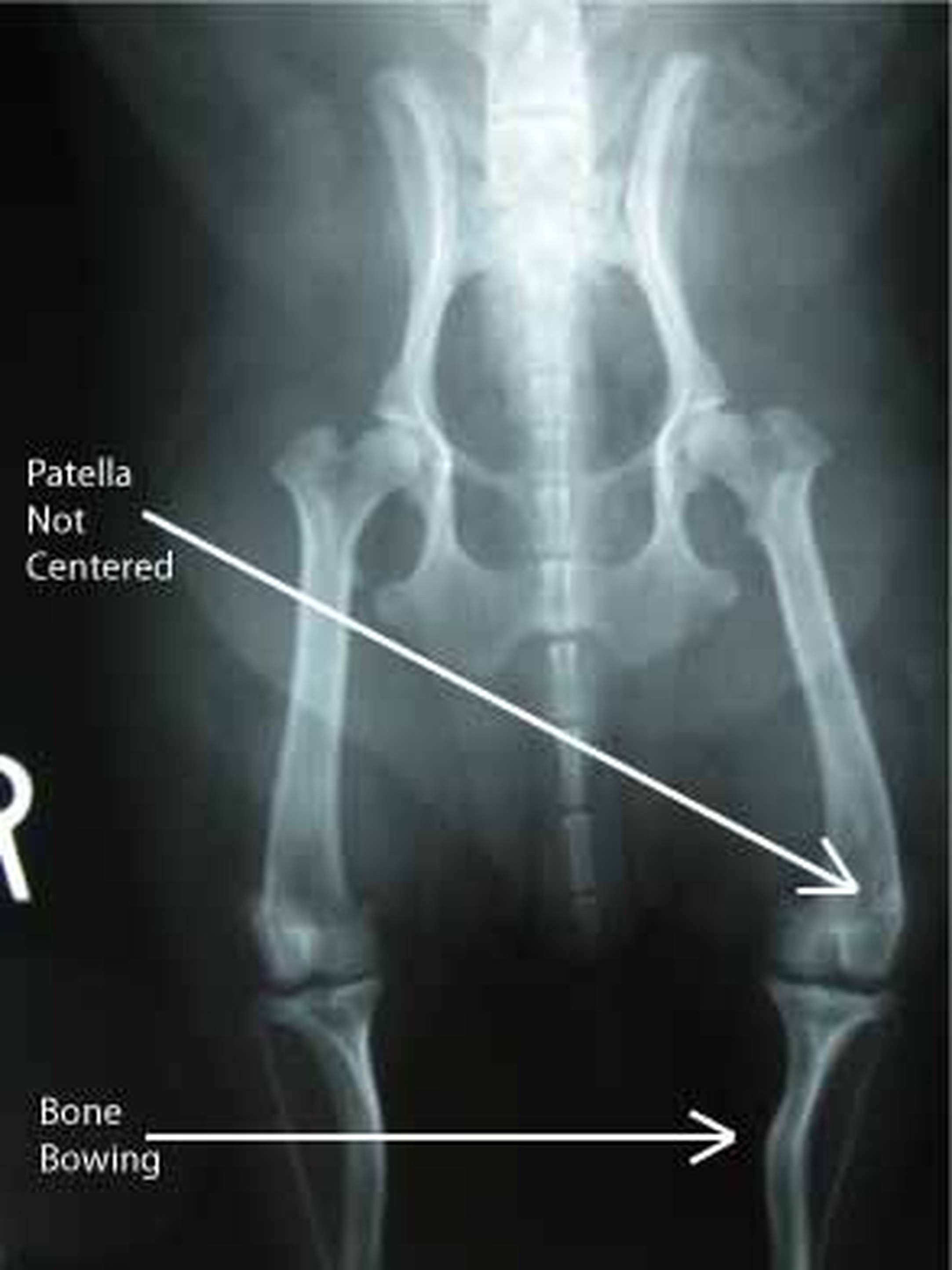
Patellar luxation occurs when the dog's kneecap (patella) is dislocated from its normalanatomic position in the groove of the thigh bone (femur). When the kneecap is dislocated from the groove of the thigh bone, it can only be returned to its normal position once the quadriceps muscles in the hind legs of the animal relax and lengthen. It is for this reason that most dogs with the condition will hold up their hind legs for a few minutes.

A dislocated kneecap is one of the most prevalent knee joint abnormalities in dogs. The condition is most common in toy and miniature dog breeds such as the Yorkshire Terrier, Dachshund, West Highland White Terrier (Westie), Pomeranian, Pekingese, Chihuahua, and Boston Terrier. Female dogs are 1 1/2 times more likely to acquire the condition.

Symptoms and Types
The specific symptoms of a dislocated kneecap will depend on the severity and persistence of the condition, as well as the amount of degenerative arthritis that is involved. Typically, a dog with a dislocated kneecap will exhibit prolonged abnormal hindlimb movement, occasional skipping or hindlimb lameness, and sudden lameness.

The dog will rarely feel pain or discomfort once the kneecap is out of position, only feeling pain at the moment the kneecap slides out of the thigh bone's ridges.

Causes
A dislocated kneecap is usually caused by a genetic malformation or trauma. The clinical signs of the condition will normally start showing approximately four months after birth.

Diagnosis
A dislocated kneecap is diagnosed through a variety of means. Top view (craniocaudal) and side view (mediolateral) X-rays of the stifle joint, hip, and hock may be used to detect bending and twisting of the thigh bone and larger bone of the lower leg. Skyline X-rays may reveal a shallow, flattened, or curved groove of the thigh bone. A fluid sample taken from the joint and an analysis of the lubricating fluid in the joint (synovial fluid) will show a small increase in mononuclear cells. It is also necessary for the veterinarian to perform an examination by touch to feel for kneecap freedom.

Treatment
Medical treatment for kneecap dislocation has very little effectiveness; surgery is the preferred treatment of choice for severe cases. Surgery can correct both the affected structures and the movement of the kneecap itself, and in 90 percent of cases, frees the dog from lameness and dysfunction.

The kneecap may be fastened on the outside of the bone to prevent it from sliding towards the inside. Alternatively, the groove of the thigh bone may be deepened so that it can better hold the kneecap.

Living and Management
Follow-up treatment after successful surgery will include leash walk exercise for one month (avoid jumping) and yearly examinations to check for progress. It is important that pet owners are aware that there is a high possibility of recurrence (48 percent), although the dislocation will be considerably less severe than the original incidence. Because kneecap dislocation is genetically inherited, the breeding of affected dogs is highly discouraged.

PreventionThere are currently no known preventative measures for this medical condition.

1 Comment
<<Previous
Forward>>
    PET SCAN Blog: 
    The INNER Pet... 
    Pet Health Inside & Out
    The goal of this blog is to help educate pet owners by sharing pet health facts and interesting pet news articles

    Archives

    September 2019
    August 2019
    June 2016
    March 2016
    November 2015
    June 2015
    April 2015
    March 2015
    January 2015
    December 2014
    November 2014
    October 2014
    September 2014
    August 2014
    July 2014
    June 2014
    May 2014
    April 2014
    March 2014
    February 2014
    January 2014

    Categories

    All
    Cancer In Pets
    Cat Health
    Dog Health
    Fun Facts
    Pets In The News
    Research News
    Videos

    RSS Feed

"YOUR Partner for Advanced Diagnostic Excellence"
Providing mobile ultrasound consultation services to veterinarians in Chandler, Gilbert, Mesa, Tempe, Ahwatukee, Scottsdale, and the Greater Phoenix Metropolitan Area. 
Picture


Proud member of the
American Veterinary Medical Association