Pet Scan:  Mobile Veterinary Ultrasound Service
  • Home
  • Indications for Ultrasound
  • Services
  • What To Expect
  • Our Doctors
  • Our Practice Partners
  • Pet Owners
  • Referring DVMs

Case Spotlight: Vomiting Cat

3/17/2016

0 Comments

 
Picture
A five-year-old neutered male Domestic Shorthair cat was presented with an acute history of anorexia and vomiting. Physical examination revealed a painful cranial abdomen. A complete blood count and blood chemistry profile were normal, and abdominal radiographs were unremarkable.

Findings
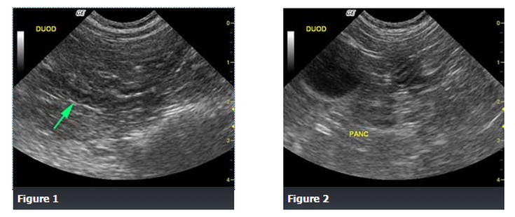
Abdominal ultrasound revealed a spastic duodenum (Figure 1), an enlarged, irregular, hypoechoic pancreatic body (Figure 2) and a hypoechoic left pancreatic lobe (Figure 3), as well as hyperechoic surrounding mesentery which surrounded the body and left pancreatic lobe. 

Differential Diagnosis
Pancreatitis
Acute gastroenteritis
Inflammatory bowel disease
Gastrointestinal obstruction
Peritonitis
Acute renal failure​

Diagnosis
Pancreatitis

Discussion
The cat responded well to medical management of pancreatitis with intravenous fluid therapy, famotidine and pain medication.

Pancreatitis is inflammation of the pancreas and it can be acute (sudden) or chronic (recurrent) in nature.  It is associated with a range of clinical signs that vary in severity, often overlapping with signs of acute gastroenteritis, inflammatory bowel disease, gastrointestinal obstruction, peritonitis, or acute renal failure.  Therefore, ultrasound is a very useful diagnostic tool to help differentiate among these similar presenting conditions.

The cause of pancreatitis in dogs and cats is usually unknown.

Source: soundvet.com
0 Comments

Case Spotlight: Older Cat with Blood in Urine

11/6/2015

0 Comments

 
Picture
This is a case is a 13-year-old female spayed domestic short haired cat with history of hematuria (blood in the urine).

Findings
​The right kidney is markedly enlarged, with a rounded shape. It is distorting the abdominal wall on the v/d projection and displacing the ascending and transverse colon medially and ventrally. The left kidney also has an abnormal shape, with reduced length and increased width.
​
There is also spondylosis deformans at the L5-6 intervertebral disc space, which is an incidental finding.
On ultrasound images, both kidneys were surrounded by large cystic structures filled with echogenic fluid. There are fine septae visible in the pseudocyst surrounding the left kidney. Drainage of both collections of fluid were clear and colorless except for the last 15 ml from the right kidney, which was hemorrhagic. Both kidneys were small and irregular with poor corticomedullary distinction.

Differential Diagnosis
  • Perinephric pseudocysts
  • Hydronephrosis
  • Chronic renal disease
  • Subcapsular hematoma
  • Lymphosarcoma

Diagnosis
Bilateral perinephric pseudocysts and chronic renal disease.


Discussion
Perinephric pseudocysts are associated with chronic renal disease, but are often asymptomatic. The association between the pseudocysts and this cat’s hematuria is unclear. The hematuria may have been due to pressure from the pseudocysts, or idiopathic in nature.

Source: veterinaryradiology.net / Allison Zwingenberger
0 Comments

Valley Fever (Coccidioidomycosis)

4/16/2015

0 Comments

 
San Joaquin Valley Fever, now called Valley Fever for short, was named because of an outbreak among farm workers in the well known San Joaquin Valley of California. The condition is caused by inhaling dust or dirt contaminated with a fungus called Coccidioides immitis. The fungus is inhaled into the lungs where it may simply cause a low grade chronic cough and calcification (mineralization) of local lymph nodes or, if the immune system is weak, it can cause pneumonia or even disseminate throughout the body.

About the Fungus

The fungus lives only in one area: the Lower Sonoran life zone. This area includes the southwest United States, Mexico and Central America as well as parts of South America. In these places, soil is sandy and alkaline. There is little rainfall. Summer is hot and winter is cold. Fungal spores, called arthroconidia, are released from the ground after a rainfall and are carried by the wind. Earthquakes or high winds or even crop harvesting have led to outbreaks of this infection as both conditions stir up fungal spores. Once the spores are in the air, they can be inhaled and set up infection.

Getting Sick

It has been estimated that 60% of animals and people that become infected with this fungus never get sick. It is only when antibodies indicating past infection show up on a blood test that infection is discovered.

But the other 40% do get sick. It only takes 1 to 3 weeks from the inhalation of fungal spores before coughing starts. Fever, weight loss, and appetite loss accompany the cough, which can be dry or productive. If the fungus escapes the lung and travels out elsewhere in the body, the infection has disseminated and prognosis is much worse. On the average, several months pass between the initial cough (which may be so mild as to have gone undetected) and signs in another organ system. There is an order by which the fungus disseminates in that it appears in certain organs before others. After the lung, the fungus spreads to bone, then eyes, heart, testicles, brain, spinal cord, and abdominal organs. In cats, the pattern is a bit different with the usual disease picture being draining skin lesions with fever, weight loss and poor appetite. The respiratory phase is rarely noted in feline patients.

An animal or person who is sick is not contagious; infection is only through inhalation of a fungal spore released from dirt, but only a few spores are necessary for infection to occur.

Testing

There are several diagnostics that can be helpful with this condition.

  • Radiographs
    The patient with a cough is likely to have x-rays taken of the chest. Findings will not be specific to Valley Fever; large lymph nodes and lung infiltration will likely be found. The point is to rule out other conditions that are more readily identifiable on radiographs such as heart disease, tracheal collapse, or more localized forms of pneumonia.
  • Cytology
    If some sort of tracheal wash or lavage is performed, where samples of lung fluid are collected for analysis, it is possible to capture some fungal spores. If this occurs, the diagnosis is confirmed, as Coccidioides immitis has a fairly characteristic large size and appearance.


  • Blood testing
    Blood testing is by far the least invasive way of diagnosing Valley Fever. Antibodies against the fungus are measured in a blood sample using one or even several different methods (tube precipitin, complement fixation, latex agglutination, gel immunodiffusion, or enzyme linked immunoassay). As mentioned, many patients are exposed to this fungus and do not get sick. These patients will absolutely have positive antibody tests so it is important not to treat an animal with a positive antibody level (also called an antibody titer) and no history of symptoms of any kind.
Treatment

A general rule with any fungal infection is that it takes a long time to clear. Valley Fever is no different and a year of medication administration is fairly common. Treatment continues until the clinical signs have resolved, the radiographs look normal, and the antibody level has stabilized (note antibody levels may never fully drop to zero). Ketoconazole, itraconazole,  and fluconazole are the usual drugs to treat this condition. Monitoring liver enzymes is important with all of these, particularly since long treatment courses are common.

Lufenuron, the active ingredient in the flea control product Program, was developed to inhibit the development of chitin (the crunchy exterior material of the insect body). The idea was that a flea larva who had fed upon blood pellets (flea dirt) from a pet treated with oral lufenuron would be unable to properly pupate into an adult flea. It turns out that the shell of Coccidioides immitis is also rich in chitin and that lufenuron at specific doses may be helpful in clearing this infection. This is still an experimental use but it may be worthwhile to ask your veterinarian about this product.

If the disease is limited to the lung, prognosis is felt to be good. The most severe form of infection appears to involve bone and when multiple bones are infected, complete recovery is unlikely unless amputation is possible. Eye involvement is painful and very difficult to clear short of removing the eye (enucleation). Obviously, a patient with any internal fungal infection should avoid immunosuppressive drugs for other conditions as their use could lead to a disastrous fungal dissemination.

Source: Veterinary Partner

0 Comments

Immune Mediated Hemolytic Anemia (IMHA)

3/20/2015

0 Comments

 
Immune-mediated hemolytic anemia is a condition where the patient’s immune system begins attacking her own red blood cells. What occurs on a microscopic level is that the branch of the immune system that produces antibodies begins to direct them against the patient’s own red blood cells. The red blood cells quickly become coated with tiny antibody proteins, essentially marking these red blood cells for destruction. When too many red blood cells are destroyed the patient is said to be anemic and will feel cold and weak. Because the red blood cells are being destroyed internally (and not lost through bleeding), the patient will become yellow-tinged (jaundiced) rather than pale (see picture below).

Removing old Red Cells/Recycling Iron: The Normal System

Red blood cells have a natural life span from the time they are released from the bone marrow to the end of their oxygen-carrying days when they become too stiff to move through the body’s narrow capillaries. A red blood cell must be supple and flexible to participate in oxygen delivery and carbon dioxide removal, so when the cell is no longer functional the body destroys it and recycles its components.

When old red blood cells circulate through the spleen, liver, and bone marrow, they are plucked from circulation and destroyed in a process called extravascular hemolysis. Their iron is sent to the liver for recycling in the form of a yellow pigment called bilirubin. The proteins inside the cell are broken down into amino acids and used for any number of things (burning as fuel, building new protein etc.) The spleen uses immunological cues on the surface of red blood cells to determine which cells are plucked out of circulation. In this way, red cells parasitized by infectious agents are also removed from circulation along with the geriatric red cells. When the immune system marks too many cells for removal, problems begin.

The jaundiced/icteric pet has a yellow color in the gums and whites of the eyes.The spleen enlarges as it finds itself processing far more damaged red blood cells than it normally does. The liver is overwhelmed by large amounts of bilirubin and the patient becomes jaundiced (icteric), which means her tissues become a yellow/orange color.

Making matters worse, a protein system called the complement system is activated by these anti-red cell antibodies. Complement proteins are able to simply rupture red blood cells if they are adequately coated with antibodies, a process called intravascular hemolysis. Ultimately, there aren’t enough red blood cells left circulating to bring adequate oxygen to the tissues and remove waste gases.  A life-threatening crisis has emerged; in fact 20-80% mortality (depending on the study) has been reported with this disease.

Signs you Notice at Home
 
The jaundiced/icteric pet has a yellow color in the gums and whites of the eyes. Illustration by Wendy Brooks.Your pet is obviously weak. She has no energy and has lost interest in food. Urine is dark orange or maybe even brown. The gums are pale or even yellow-tinged as are the whites of the eyes. There may be a fever. You (hopefully) brought your pet to the veterinarian’s office as soon as it was clear that there was something wrong.
 
The Tests Show Anemia

Part of a general evaluation includes blood testing. If your pet seemed obviously pale or jaundiced (yellow-tinged), right off the bat your vet may run a test called a packed cell volume. This test can be run in most veterinary clinics and involves spinning a small amount of blood in a small glass tube at high speed to separate the red blood cells from the serum (the blood’s liquid phase). By comparing the blood tube to a chart, it will become obvious if your pet has a low red blood cell count. If your pet has hemolytic anemia, the serum will be bright orange instead of its normal off-white color.

The anemia may be seen on a full blood panel sent to a reference laboratory. These laboratories perform a test called a hematocrit, which is slightly different from a packed cell volume but essentially measures the same thing: the percentage of blood volume made up by red blood cells. This number should be 43 - 59 for dogs and 29 - 50 for cats.

Anemia is the condition where one’s red blood cell count is low. Anemia can be mild or severe and can represent bleeding, red blood cell destruction (as in IMHA), or simply lack of red blood cell production. Once a patient is found to be anemic, it is important to determine why.

The Tests Show Responsive Anemia

Anemia due to poor red blood cell production by the bone marrow is called a non-responsive anemia. Such anemias are caused by chronic inflammatory diseases (like inflamed skin, infected teeth, or other long standing irritations), kidney failure, cancers of various types, or certain drugs (especially agents of chemotherapy).

Normally when red blood cells are lost, the drop in blood oxygen that results causes hormonal changes leading to increased production of red blood cells by the bone marrow. These are called responsive anemias because the bone marrow is responding. Bleeding and immune mediated red blood cell destruction are both responsive anemias.

There are several ways to determine if the anemia is responsive or not from the blood panel results. Most blood panels run by reference laboratories include a portion called a complete blood count, or CBC, which reviews red blood cell count, size, shape, and maturity as well as white blood cell types and ratios. A patient with a responsive anemia will have an active bone marrow. Red blood cells will be released early leading to a variety of sizes and redness of red blood cells circulating in the blood (less mature red blood cells are larger and paler than mature cells). Further, red blood cell precursors called reticulocytes are released. (These may be thought of as red blood cells so immature they can’t truly be called red blood cells yet.) If the bone marrow stimulus is especially strong, red blood cells may be released still containing cell nuclei.

These findings indicate the anemia is responsive. This means either red blood cells are being lost through bleeding, possibly internal bleeding, or they are being destroyed by the immune system.

Which is it?

The Tests Suggest Immune-Mediated Destruction rather than Bleeding

There are several clues in blood testing that tell us if our patient is bleeding or destroying red blood cells. You might think it would be obvious if the patient were bleeding but if the bleeding is internal it may not be so obvious.

Icterus (also Called Jaundice)
We have mentioned this but let’s recap. Icterus is the yellow color that is taken up by a patient’s tissues when the liver is overwhelmed with bilirubin, the iron-containing by-product of red blood cell destruction.  Normally red blood cells are removed from the circulation when they become old and inflexible. Their iron is recycled in the liver. With so many red cells being destroyed, the liver is overwhelmed and bilirubin (a yellow pigment) spills out everywhere, coloring urine, gums, skin, and the eyes orange.

Is immune-mediated red cell destruction the only cause of icterus? Absolutely not. Liver failure also leads to icterus when the diseased liver cannot process normal amounts of bilirubin. In cats especially, bacterial endotoxin (the toxic cell walls of certain types of infecting bacteria) can lead to icterus. Usually, however, a responsive anemia together with icterus, suggests immune-mediated red cell destruction.

Spherocytes

A normal red blood cell is concave on both sides and shaped like a disc. It is slightly paler in the middle than on its rim. After a portion has been bitten off, it re-shapes into a more spherical shape with a denser red color. The presence of spherocytes indicates that red blood cells are being destroyed.

Autoagglutination
In severe cases of immune-mediated hemolytic anemia, the immune destruction of red cells is so blatant that the red cells clump together (because their antibody coatings stick together) when a drop of blood is placed on a microscope slide. Imagine a drop of blood forming not a red spot but a yellow spot with a small red clump inside it. This finding is especially foreboding.

Leukemoid Reaction
Classically, in IMHA the stimulation of the bone marrow is so strong that even the white blood cells lines (which have little to do with this disease but which also are born and incubate in the bone marrow alongside the red blood cells) are stimulated. This leads to white blood cell counts that are spectacularly high.

More Tests Needed

Coomb’s Test (also Called a Direct Antibody Test)
This test is designed to identify the antibodies that are coating red blood cell surfaces. This test is the current state of the art for the diagnosis of IMHA but, unfortunately, it is not as helpful as it might seem. It can be erroneously positive if there is any inflammation or infectious disease (which might lead to harmless attachment of antibody to red cell surfaces) or in the event of a prior blood transfusion (ultimately transfused red cells are removed from the immune system). The Coomb’s test can be erroneously negative for a number of reasons as well. If the clinical picture fits with IMHA, often the Coomb’s test is skipped.

Remember, not all causes of hemolysis (red blood cell destruction) are immune-mediated.  Onions in large amounts, and possibly garlic, will cause a toxic hemolysis. Zinc toxicity, usually caused from swallowing a penny minted after 1983 or from licking a zinc oxide ointment applied to the skin, will cause hemolysis as well. In a young animal, a genetic red blood cell malformation might be suspected.

Once there is a diagnosis of IMHA, efforts to determine an underlying cause should be made. 

Treatment and Monitoring During the Crisis

The patient with IMHA is often unstable. If the hematocrit has dropped to a dangerously low level, then blood transfusion is needed. It is not unusual for a severely affected patient to require many transfusions. General supportive care is needed to maintain the patient’s fluid balance and nutritional needs. Most importantly, the hemolysis must be stopped by suppressing the immune system’s rampant red blood cell destruction. We will review these aspects of therapy.

Transfusion
There are several products that may be helpful in treating the IMHA case. If the patient is in a crisis and needs immediate therapy, artificial blood may be a good choice. Artificial blood (Oxyglobin®) is made from hemoglobin harvested from cow’s blood. Because the patient does not receive actual red blood cells, the artificial blood does not further stimulate the immune system. Artificial blood does not require refrigeration and is likely sitting on the shelf ready to use at your veterinarian’s office. The disadvantage of artificial blood is that it does not last in the body like a well-matched blood transfusion does. The body begins removing artificial blood immediately so that the entire transfusion is probably gone in 48 hours or so. In IMHA, this may buy some time but since IMHA tends to have a long treatment course, it is likely that the patient will be back where they started from at that point. If a compatible donor is not readily available, sometimes an artificial blood transfusion buys enough time to find a compatible donor

Well-matched whole blood or packed red cells (a unit of whole blood with most of the plasma, leaving only a concentrated solution of red blood cells) may last longer. Compatible blood can last a good 3 to 4 weeks in the recipient’s body. The problem, of course, with IMHA is that even the patient’s own red blood cells are being destroyed so what chance do donated cells have? Cross matching of red cells is ideal but still may not lead to a good match given the hyperactivity of the patient’s immune response. For this reason, it is not unusual for several transfusions to become necessary during treatment.

Immune Suppression
Corticosteroid hormones in high doses are the cornerstone of immune suppression. Prednisone and dexamethasone are the most popular medications selected. These hormones are directly toxic to lymphocytes, the cells that produce antibodies. If the patient’s red blood cells are not coated with antibodies, they will not have been targeted for removal so stopping antibody production is an important part of therapy. These hormones also suppress the activity of the reticuloendothelial cells that are responsible for removing antibody-coated red cells.

Corticosteroids may well be the only immune suppressive medications the patient needs. The problem is that if they are withdrawn too soon, the hemolysis will begin all over again. The patient is likely to be on high doses of corticosteroids for weeks or months before the dose is tapered down and there will be regular monitoring blood tests. Expect your pet to require steroid therapy for some 4 months; many patients must always be on a low dose to prevent recurrence.

Corticosteroids in high doses produce excessive thirst, re-distribution of body fat, thin skin, panting, predisposition for urinary tract infection and other signs that constitute Cushing’s Syndrome. This is an unfortunate consequence of long-term steroid use but in the case of IMHA, there is no way around it. It is important to remember that the undesirable steroid effects will diminish as the dosage diminishes.

More Immune Suppression

If no response at all is seen with corticosteroids, supplementation with stronger immune suppressive agents is necessary. The two most common medications used in this case areazathioprine and cyclophosphamide. These are serious drugs reserved for serious diseases. Follow the links above to read more about specific side effects, concerns, etc.

Cyclosporine is an immune-modulator made popular in organ transplantation technology. It has the advantage over the two above medications of not being suppressive to the bone marrow cells. It has been a promising adjunctive therapy in IMHA but has two major problems: first, it is extremely expensive and second, blood level monitoring is necessary to ensure that the dosage is appropriate. This adds dramatically to the expense of treatment but ultimately may provide results not possible with other drugs.

Leflunomide is an immuno-modulator that is meant for patients with immune mediated diseases when corticosteroids either do not work or cannot be used. It is expensive (approximately $600 per month) but we may be hearing more about it in the future.

Human gamma globulin transfusion is a treatment that is reserved for patients for whom more traditional methods of immune suppression have been ineffective. The gamma globulin portion of blood proteins includes circulating antibodies. These antibodies bind the reticulo-endothelial cell receptors that would normally bind antibody-coated red blood cells. This prevents the antibody-coated red blood cells from being removed from the circulation. Human gamma globulin therapy seems to improve short-term survival in a crisis but, unfortunately, its availability is limited and it is very expensive.

Why did this Happen to your Pet?

When something as threatening as a major disease emerges, it is natural to ask why it occurred. Unfortunately, if the patient is a dog, there is a good chance that there will be no answer to this question. Depending on which study you read, 60-75% of IMHA cases do not have apparent causes.

In some cases, though, there is an underlying problem: something that triggered the reaction. A drug can induce a reaction that stimulates the immune system and ultimately mimics some sort of red blood cell membrane protein. Not only will the immune system seek the drug but it will seek proteins that closely resemble the drug and innocent red blood cells will be consequently destroyed. Drugs most commonly implicated include penicillins, trimethoprim-sulfa, and methimazole.

Drugs are not the only such stimuli; cancers can stimulate exactly the same reaction (especially hemangiosarcoma).

Red blood cell parasites create a similar situation except the immune system is aiming to destroy infected red blood cells. The problem is that it gets over-stimulated and begins attacking the normal cells as well.

There is some thinking that vaccination can trigger IMHA. Insect bites have also been implicated. Both have been temporally associated with the development of autoimmune hemolytic anemia. The relationship between recent vaccination and IMHA development is one of the factors that has led most universities to vaccinate dogs with the standard DHLPP every three years rather than annually.

Dog breeds predisposed to the development of IMHA include: cocker spaniels, poodles, Old English Sheepdogs, and Irish setters.

In cats, IMHA generally has one of two origins: feline leukemia virus infection or infection with a red blood cell parasite called Mycoplasma hemofelis (previously known as Hemobartonella felis).

Complications of IMHA

Thromboembolic Disease
This particular complication is the leading cause of death for dogs with IMHA (between 30-80% of dogs that die of IMHA do so due to thromboembolic disease). A thrombus is a large blood clot that obstructs (occludes) a blood vessel. The vessel is said to be thrombosed. Embolism refers to smaller blood clots spitting off the surface of a larger thrombus. These mini-clots travel and obstruct smaller vessels, thus interfering with circulation. The inflammatory reaction that normally ensues to dissolve errant blood clots can be disastrous if the embolic events are occurring throughout the body.

Heparin, a natural anticoagulant, may be used as a preventive in hospitalized patients or in patients with predisposing factors for embolism.

*Four to seven days are required for the bone marrow to generate a response. If hemolysis occurs suddenly there may not have been adequate time for a response. When this occurs, if there is any question about the responsive nature of the anemia, continued monitoring of the complete blood count will show a clear response in an appropriate time period. It should also be noted that in an especially unlucky patient, the red blood cell destruction may extend to the pre-red blood cells (reticulocytes, nucleated red cells and other precursors) within the bone marrow. If these cells are also destroyed, the condition is especially dangerous and it will take weeks rather than days to begin to see a response to treatment. The lack of circulating immature red cells will lead this anemia to test as non-responsive.

The 2002 study by Drs. Anthony Carr, David Panciera, and Linda Kidd at the University of Wisconsin School of Veterinary Medicine reviewed 72 dogs with IMHA looking for trends. Their findings are:

  • The only predisposed breed they found was the cocker spaniel.  
  • Most patients were female.  
  • The mean age was 6.8 years.  
  • Timing of vaccination was not associated with the development of IMHA.  
  • 94% of cases had spherocytes on their blood smears. 
  • 42% showed autoagglutination.  
  • 70% also had low platelet counts.  
  • 77% were Direct Coombs' positive.  
  • 58% were suspected of having disseminated intravascular coagulation.  
  • 55% required at least one blood transfusion.  
  • Mortality rate was 58%.  
  • Of those that died, 80% had thromboembolism present on necropsy (autopsy).

Prognostic Factors for Mortality and Thromboembolism in Canine Immune-Mediated Hemolytic Anemia. A.P. Carr, D. Panciera, L. Kidd. Journal of Veterinary Internal Medicine. 2002; 16: 504-509.

Another study
The 2005 study looking for trends by Drs. Tristan Weinkle, Sharon Center, John Randolph, Stephen Barr, and Hollis Erb at Cornell University reviewed 151 dogs with IMHA. They found:

  • Cockers spaniels and miniature schnauzers were both overrepresented (i.e., felt to be predisposed). These breeds, however, showed the same mortality rate as other breeds.  
  • Unspayed female dogs were overrepresented.  
  • Neutered male dogs were more commonly affected than unneutered male dogs (begging the question of whether male hormones might have some protective effect).  
  • The chance of survival either long term or short term was significantly enhanced by the addition of aspirin to the treatment protocol, especially when combined with azathioprine.  
  • Adequate vaccination information was not obtained for enough patients to comment on association with vaccination.  
  • 89% of affected dogs showed spherocytes on their blood smears.  
  • 78% showed autoagglutination.  
  • 70% of patients required at least one blood transfusion.  
  • Of the 151 dogs studied, 76% survived, 9% died, and 15% were euthanized. Survivors were hospitalized an average of 6 days. Non-survivors were hospitalized an average of 4 days.  
  • 100% of dogs that died or were euthanized showed thromboembolism on necropsy (autopsy).  
  • Of the dogs that survived 60 days or more, 15% experienced relapse. Most dogs treated with corticosteroids, azathioprine, and ultra-low dose aspirin did not experience relapse.

Source: Veterinary Partner
0 Comments

High Blood Pressure in Cats

1/30/2015

0 Comments

 
Systemic Hypertension in Cats
Hypertension, more commonly referred to as high blood pressure, occurs when the cat's arterial blood pressure is continually higher than normal. When it is caused by another disease, it is called secondary hypertension; primary hypertension, meanwhile, refers to when it actually is the disease. Hypertension may affect many of the cat's body systems, including heart, kidneys, eyes, and the nervous system.

Symptoms and Types
The following are just some of the more common symptoms displayed by cats with high blood pressure:

  • Seizures
  • Circling
  • Disorientation
  • Blindness
  • Dilated pupils
  • Retinal detachment
  • Hemorrhage of the eye
  • Blood in the urine
  • Protein in the urine
  • Bleeding from the nose
  • Swollen or shrunken kidneys
  • Heart murmurs
  • Weakness, either on one side of the body or in the legs
  • Involuntary oscillation (rolling) of the eyeballs
  • Palpable thyroid gland (when hyperthyroid)

Causes

The cause of primary hypertension in cats is not known. However, it may have a genetic component. So how prevalent is this form of hypertension? Studies have varied, but one study found that 65 percent of cats with chronic renal failure and 87 percent of cats with hyperthyroidism had mild high blood pressure. Ages of cats with hypertension ranged 4 to 20 years old.

Secondary hypertension, which accounts for 80 percent of all hypertension cases, may be due to a variety of factors, including renal disease, hormonal fluctuation, and hyperthyroidism.

Diabetes may also be a cause for hypertension, although it is uncommon in cats. If you suspect that your cat is suffering from hypertension, bring it in so that your veterinarian may provide a proper diagnosis.

Diagnosis
Blood pressure is often measured in pets in the same manner as in humans. An inflatable cuff will be placed on the cat's paw or tail, and standard blood pressure measuring instruments will check the pressure. It is important to keep the cat still long enough to get an accurate reading.

The standards for cat blood pressure are:

  • 150/95 – at this reading or below, there is minimal risk and treatment is not recommended
  • 150/99 to 159/95 -- intervention is rotuinely not recommended at these readings
  • 160/119 to 179/100 -- treatment should be sought to limit the risk of organ damage
  • 180/120 -- immediate treatment should be sought to limit the degree of other more severe complications

Five to seven measurements are generally taken. The first measurement will be discarded, and the cat's excitement level during the procedure will be taken in account. If the results are in dispute, the procedure will need to be repeated.

Treatment
The underlying cause of the high blood pressure will be treated first. Otherwise, the cat will probably be on medication to control the blood pressure indefinitely. The medication of choice is either a calcium channel blocker or a beta-blocker. As to cat's diet, the veterinarian may recommend food that are lower in sodium.

Blood pressure should be checked regularly, and some lab tests may be ordered by your veterinarian to measure your cat’s reactions to the medication.
Source: PetMd
0 Comments

New feline kidney disease research offers hope for earlier detection and improved quality of life

1/30/2015

0 Comments

 
Foundation For Feline Renal Research, the only 501(c)(3) nonprofit foundation dedicated exclusively to the understanding and treatment of feline kidney disease, is pleased to announce a $20,000.00 grant to the Royal Veterinary College, London, in support of their groundbreaking study "Unravelling The Genetic Basis for Blood Pressure and Kidney Function In the Cat". This important work, which is starting now, will be led by Dr. Rosanne Jepson, BVSc (Dist) MVetMed PhD DipACVIM DipECVIM MRCVS and Dr. Jonathan Elliott, MA, Vet MB, PhD, Cert SAC, Dip ECVPT, MRCVS.

Kidney disease is one of the biggest killers of companion cats; it causes great pain and suffering, and there is no cure. As many as one in three cats will ultimately develop the disease. The goal of this study is to isolate and analyze genes that influence renal disease and hypertension (the two diseases often go hand in hand).

According to Dr. Jepson, "Kidney disease is one of the most common conditions that we see in ageing cats. Some of these cats also develop high blood pressure (hypertension) which can result in damage, particularly to their eyes, and can lead to blindness. We know that, in human medicine, there are genes that are involved in both kidney disease and hypertension. At the Royal Veterinary College we have been studying these conditions for over 20 years. Thanks to the funding provided by Foundation For Feline Renal Research, we are now in a position to use newly developed cutting edge technology to look for gene associations in almost 1000 cats. As far as we know, this will be the first and largest study looking at genes associated with kidney disease and blood pressure in older cats. It's really exciting to be starting new work that we hope will expand our knowledge of both of these important conditions."

This humane study utilizes DNA from blood samples obtained for diagnostic purposes from companion cats during the course of their regularly scheduled veterinary visits. The samples, after being used for the necessary diagnostic purposes, were preserved for use in this study with the consent of the cats' human companions.

0 Comments

Using Viruses to Treat Cancer in Pets

12/12/2014

0 Comments

 
Surgery, radiation, and chemotherapy are the more commonly known treatments for cancer in pets. But newer technologies are opening up other possibilities. A recent experiment summary (abstract) suggests the use of genetic modified viruses to treat various types of cancer.

Oncolytic Virotherapy
The idea of using a virus for cancer treatment or oncolytic virotherapy is not a new idea. In the 1940s scientists conducted animal studies using viruses to treat tumors. Doctors in the 1950s observed that cancer patients that were stricken with viral infections or recently vaccinated experienced improvement in their condition. It was believed that the infections or vaccinations triggered an immune response that increased the production of interferon and tumor necrosis factors, or TNFs.

Interferons are large molecules released by cells infected with viruses, bacteria, parasites and tumors to interfere, hence their name, with virus reproduction and to trigger responses from immune cells. Interferons activate natural killer white blood cells and large white cells called macrophages that attack and destroy the invading organisms and cancer cells. Interferon promotes the production or molecular complexes that attach to viral, bacterial, parasite, and tumor cells so they are more quickly and effectively attacked by killer white cells. TNFs causes destructive changes in cell walls and causes foreign or tumor cells to burst and die

Despite the potential for viral therapy of cancer in these early years, it required the present advances in technology to achieve a real possibility. Precisely, it required our present ability to genetically modify organisms like viruses and safely use them to target cancer cells. The viruses are modified to prevent their normal ability to cause disease and genetically altered to produce interferon or other anti-cancer molecules.

Preliminary Study in Dogs
The abstract was based on a small study intended to evaluate the safety and effectiveness of a new oncolytic virus. The group was made up of of seven dogs suffering from various cancers (lymphoma, malignant melanoma and multiple myeloma). The researchers used a novel virus for their study; they used a modified vesicular stomatitis virus that causes oral, udder, and hoof ulcers in cattle. Although seldom fatal, the disease causes inappetence and decreased milk or meat production [in cattle]. It can also infect horses and pigs, and rarely, sheep, goats, and llamas. Because of its effect on agricultural production, vesicular stomatitis is a diagnosis that requires mandatory reporting to federal and state animal health officials.

The virus was also modified to produce human or canine interferon. Three dogs received the human form and four dogs received the canine form. The abstract reported measurable improvement but did not specify the type and extent of the improvements except for the production of neutralizing antibodies within 7-10 days after viral administration. Side effects were minimal and included reversible changes in liver enzymes, fever, and urinary tract infection. Virus was not shed in the urine or saliva. These limited side effects are comparable or even fewer than those expected with radiation or chemotherapy.

This is a small study and rightly titled as preliminary. It has yet to be published so critical evaluation is still not available. Clearly, much more study is required for this type of treatment. What is exciting is that this is one of many new potential treatments for cancer in pets. Advanced cancer treatment in the last decade has changed how the diagnosis is now viewed. Rather than an immediate death sentence, cancer can now be better managed as a chronic disease much like kidney and heart conditions. These new treatments offer greater treatment flexibility and potentially an improved quality of life.

0 Comments

Bladder Inflammation in Cats

10/8/2014

0 Comments

 
Picture


Interstitial Cystitis in Cats, Feline Interstitial Cystitis (FIC)

Feline interstitial cystitis, sometimes called feline idiopathic cystitis or FIC, is an inflammation of the bladder that causes symptoms of lower urinary tract disease. However, in the case of interstitial cystitis, a definitive cause for the disease cannot be identified.

Feline interstitial cystitis can occur in both female and male cats. It is a chronic disease that can be difficult to treat and frustrating for cats and cat owners alike.

Symptoms and Types
Symptoms associated with interstitial cystitis include:

  • Frequent attempts to urinate
  • Straining to urinate
  • Urinating in inappropriate places in the house
  • Crying out during attempts to urinate
  • Blood-tinged urine

Causes

Though the cause of feline interstitial cystitis is not fully understood, stress and the changes inherent in the body as a result of stress are thought to play a large part in interstitial cystitis. Some researchers believe that interstitial cystitis is only one of the manifestations seen in cats suffering from stress and may only be the “tip of iceberg” in terms of symptoms that may be caused by stress. Abnormalities have also been found in the nervous, endocrine and cardiovascular systems of cats in addition to the urinary system. It remains unknown why some cats develop symptoms of FIC and others do not.

Diagnosis
Diagnosis relies on ruling out other diseases that may cause similar symptoms, such as urinary tract infections, bladder stones, and other bladder abnormalities in cats. Testing that is frequently performed include:

  • A blood screen, including a complete blood cell count (which examines the different types of cells circulating in the blood stream, such as red blood cells and white blood cells) and chemistry profile (which is useful in evaluating the function of major organs such as the liver and kidneys)
  • A urinalysis, which checks for abnormalities in the urine, including blood, crystals, protein and other abnormal substances as well as testing the pH (which determines how acidic the urine is),  and the urine specific gravity (which determines whether the urine is concentrated or not)
  • An abdominal X-ray and/or an ultrasound exam of the bladder to rule out stones and other abnormal structures in the bladder
TreatmentTreatment consists primarily of modifying the environment to help reduce stress levels, dietary modifications, medications for pain, and other pharmaceuticals that may alter your cat’s mental state.

Multi-modal environmental modifications (MEMO) is the term that is used to describe altering the cat’s environment in an attempt to reduce the cat’s stress level. See the Living and Management section below for more information about MEMO.

Pain medications are often used in treating interstitial cystitis to relieve the discomfort caused by the inflammation within the bladder and urinary tract and make your cat more comfortable while urinating.

Dietary modifications are often necessary and you should follow your veterinarian’s directions if a special diet is recommended. Increasing water consumption is an important part of treatment and feeding canned food, when possible, will help increase the moisture content in your cat’s diet.

Pheromones such as Feliway are often recommended to help reduce stress levels for your cat.

Other medications that your veterinarian may advise include amitriptyline, clomipramine or fluoxetine, all of which are antidepressants. These medications are generally reserved for cases where MEMO, dietary modifications and pain medications have failed to help.

Living and Management
MEMO involves providing for all of your cat’s basic needs.

Litter box management is a necessary part of environmental modification.

  • Provide an adequate number of litter boxes. There should be one more litter box than the number of cats in the household.
  • Most cats prefer a large litter box over a smaller one.
  • Make sure the sides of the litter boxes are not too high for your cat. This is especially important for older cats that may suffer from arthritis or other mobility issues and for young kittens.
  • Choose an appropriate litter for your cat. The ideal litter is dust-free and fragrance-free. Strongly scented litters may be pleasant for you but not for your cat. You may have experiment with different types of litters to find the one your cat prefers.
  • Be sure to clean the litter boxes frequently.
  • Place all litter boxes in a quiet location where your cat will not be disturbed or frightened when using the box.
Provide feeding and water stations that are accessible for all cats in your household. If you have more than one cat, you may need to provide more than one feeding and water station.

Cats prefer perches, preferably at eye-level or above, on which to rest and observe their surroundings. Be sure to provide an adequate number of perches for all the cats in your household. Consider placing one or more of these perches near a window for your cat’s enjoyment.

All cats need hiding places. Your cat should have a place to retreat where he will not be disturbed by people or by other pets. In a multi-cat household, be sure there is an adequate number of hiding places for all cats.

Provide interactive toys for your cat. Food puzzles are also helpful to provide distraction for your cat and also provide exercise.


0 Comments

Aspirin Poisoning in Cats

10/8/2014

0 Comments

 
Aspirin Toxicity in Cats
Aspirin, a non-steroidal anti-inflammatory medication, has been found to have beneficial effects for some animals. It has been used for conditions related to blood clotting, inflammation, and for its analgesic properties. However, it can also be toxic to the body. Once ingested, aspirin forms salicylic acid, which is then distributed throughout the body. Aspirin toxicity is a particular concern in cats because they lack the enzyme critical for metabolizing salicylic acid properly. Cat owners must follow their veterinarian's orders strictly if aspirin is prescribed for any reason.

Symptoms and Types
The progression of symptoms can occur quickly. One of the first noticeable signs is loss of appetite. Other signs include vomiting, diarrhea, and intestinal hemorrhagebrought on by ulceration in the stomach and small intestines. The central nervous system may also be affected, causing your cat to have trouble walking, appear weak and uncoordinated, or even collapse. Loss of consciousness and sudden death can also occur.

Since even nontoxic levels can produce these symptoms, You will need to monitor your cat for any digestive problems or changes in behavior when giving your cat aspirin for any medical reason. If a significant amount of aspirin is ingested, emergency medical treatment will be necessary.

Diagnosis
If you know, or even suspect that your cat has ingested aspirin, and your cat is showing apparent symptoms of toxicity, diagnostic tests should focus on the severity of the toxicity. A complete blood profile will be conducted, including a chemical blood profile, a complete blood count, and a urinalysis. Usually an affected cat will be anemic, with electrolyte abnormalities, in addition to showing a reduction in the blood's ability to clot properly.

Treatment
Cats treated within 12 hours of ingestion, and which are presenting limited signs of distress, can have the concentration of aspirin in the body decreased through a prescribed treatment of decontamination. The sooner this care begins, the better. Your veterinarian may recommend that you decrease the amount of aspirin in the body by inducing vomiting at home before coming to the clinic, or the vomiting may be induce in the clinic. By inducing vomiting, or pumping the stomach (gastric lavage), your veterinarian will be able to remove as much aspirin as possible, lowering the chances of permanent injury. Activated charcoal may be given after vomiting to absorb some of the remaining aspirin.

Medications to encourage healing, or to protect the gastrointestinal lining are also generally prescribed. Depending on your cat's status, fluids and other supportive treatments may also be necessary. Hospitalization and repeated blood analysis will often be standard until your cat is stable.

Living and Management
Aspirin has several clinical uses. It can be prescribed as a pain reliever, an anti-inflammatory, an anti-platelet blood thinning agent, and for lowering an abnormal body temperature. If aspirin is being used for a chronic condition, such as for preventing a blockage of the blood vessel (arterial thromboembolism), it is important to follow your veterinarian’s directions. Reducing or discontinuing the aspirin dosage may be necessary if your pet is showing a susceptibility to toxicity.

0 Comments

FIV the Feline AIDS in Cats

8/13/2014

0 Comments

 
FELINE IMMUNODEFICIENCY VIRUS INFECTION (FIV) IN CATS
The feline immunodeficiency virus (FIV) infection is a complex retrovirus that causes immunodeficiency disease in domestic cats. Immunodeficiency is the medical term used to describe the body’s inability to develop a normal immune response. FIV is slow moving, capable of lying dormant in the body before causing symptoms (lentivirus). It is in the same class of viruses as the human immunodeficiency virus (HIV), the causative agent of acquired immunodeficiency syndrome (AIDS) in people.

There is no genetic susceptibility for infection, although genetics may play a role in the progression and severity of the disease. The average age is five years at the time of diagnosis, and the likelihood of infection increases with age. FIV is a transmissible disease that occurs more often in males because of their tendency to be more aggressive, and because they are more likely to roam, thereby increasing their exposure to the virus.

SYMPTOMS
  • Diverse symptoms owing to the decreased ability to develop a normal immune response. Associated immunodeficiencies cannot be distinguished clinically from feline leukemia virus (FeLV)
  • Recurrent minor illnesses, especially with upper respiratory and gastrointestinal signs
  • Mild to moderately enlarged lymph nodes
  • Inflammation of the gums of the mouth and/or the tissues surrounding and supporting the teeth is seen in 25 percent to 50 percent of cases
  • Upper respiratory tract disease is seen in 30 percent of cases - inflammation of the nose; inflammation of the moist tissues of the eye; inflammation of the cornea (the clear part of the eye, located in the front of the eyeball); often associated with feline herpes virus and calicivirus infections
  • Eye disease - inflammation of the front part of the eye, including the iris; disease of the eye in which the pressure within the eye is increased (glaucoma)
  • Long-term (chronic) kidney insufficiency
  • Persistent diarrhea seen in 10 percent to 20 percent of cases
  • Long-term, nonresponsive, or recurrent infections of the external ear and skin resulting from bacterial or fungal infections
  • Fever and wasting - especially in later stage
  • Cancer (such as lymphoma, a type of cancer that develops from lymphoid tissue, including lymphocytes, a type of white-blood cell formed in lymphatic tissues throughout the body)
  • Nervous system abnormalities - disruption of normal sleep patterns; behavioral changes (such as pacing and aggression); changes in vision and hearing; disorders usually affecting the nerves in the legs and paws.
CAUSES
  • Cat-to-cat transmission; usually through bite wounds and scratches
  • Occasional transmission of the virus at the time of birth
  • Sexual transmission is uncommon, although FIV has been detected in semen

DIAGNOSIS
Your veterinarian will perform a thorough physical exam on your cat, taking into account the background history of symptoms and possible incidents that might have precipitated this condition. A complete blood profile will be conducted, including a chemical blood profile, a complete blood count, and a urinalysis. Your doctor will need to rule out bacterial, viral, or fungal infections, and will also test for parasites and tumors before settling on a final diagnosis.


TREATMENT
Unless your cat is severely dehydrated, it will be treated on an outpatient basis. Your veterinarian will first work to manage any secondary infections. While secondary infections will not usually cause disease, your cat’s weakened immune system will given them entrée and they will cause further complications in your cat’s overall health. Surgery may be necessary for dealing with infected teeth and for the removal of tumors. A special diet plan may also need to be put into place.

LIVING AND MANAGEMENT
How much monitoring your cat will need from you depends on secondary infections and other manifestations of the disease. You will need to watch for the occurrence of infections in your sick cat, and be aware that wasting may occur, and that your pet may die of this disease. But, in general, the earlier FIV is detected, the better your cat’s chances are for living a long and relatively healthy life.

Within 4.5 to 6 years after the time of infections, about 20 percent of cats die; however, over 50 percent will remain without clinical signs of the disease. In the late stages of the disease, when wasting and frequent infections are most likely to occur, life expectancy is less than a year. Inflammation of the gums and mouth may not respond to treatment or may be difficult to treat.

In order to prevent this disease from occurring in the first place, you should vaccinate your cat against the virus, and protect your cat from coming into contact with cats that are FIV positive. You will also want toquarantine and test new cats that are coming into your household until you are sure that they are free of the virus. It is important to note that some cats will test positive for FIV if they are carriers, although they may never have symptoms of the virus, and that cats that have been vaccinated against the virus will test positive for it even though they do not carry it. Euthanasia is not normally called for when a cat has tested positive in part because of these reasons. If your cat has tested positive you will need to talk to your veterinarian about what to do to prevent possible transmission to other cats, and what symptoms to be watchful for, should they occur.

UPDATE Information: Feline Immunodeficiency Virus: Does it Really Cause Disease? (AVMA Conference, 2014)

0 Comments
<<Previous
    PET SCAN Blog: 
    The INNER Pet... 
    Pet Health Inside & Out
    The goal of this blog is to help educate pet owners by sharing pet health facts and interesting pet news articles

    Archives

    September 2019
    August 2019
    June 2016
    March 2016
    November 2015
    June 2015
    April 2015
    March 2015
    January 2015
    December 2014
    November 2014
    October 2014
    September 2014
    August 2014
    July 2014
    June 2014
    May 2014
    April 2014
    March 2014
    February 2014
    January 2014

    Categories

    All
    Cancer In Pets
    Cat Health
    Dog Health
    Fun Facts
    Pets In The News
    Research News
    Videos

    RSS Feed

"YOUR Partner for Advanced Diagnostic Excellence"
Providing mobile ultrasound consultation services to veterinarians in Chandler, Gilbert, Mesa, Tempe, Ahwatukee, Scottsdale, and the Greater Phoenix Metropolitan Area. 
Picture


Proud member of the
American Veterinary Medical Association