Pet Scan:  Mobile Veterinary Ultrasound Service
  • Home
  • Indications for Ultrasound
  • Services
  • What To Expect
  • Our Doctors
  • Our Practice Partners
  • Pet Owners
  • Referring DVMs

Dry Eye Syndrome (KCS) in Dogs

3/31/2014

1 Comment

 
Picture


Keratoconjunctivitis sicca in Dogs
Sometimes called dry eye syndrome, Keratoconjunctivitis sicca (KCS) is characterized by a deficiency of aqueous tear film over the surface of the eye and in the lining of the lids. The result is severe drying and inflammation of the cornea (the transparent front part of the eye) and conjunctiva (the clear membrane that covers the sclera -- the white part of the eye).


This condition is relatively common in dogs, particularly cocker spaniels, bulldogs, Dachshund, West Highland White Terrier (Westie), Lhasa apsos, and shih-tzus. In addition, there is some suspicion that females may be more predisposed to KCS than males.
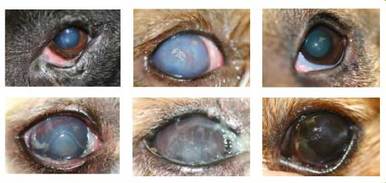
Symptoms and Types
  • Excessive blinking
  • Swollen conjunctival blood vessels
  • Chemosis (swelling of the tissue that lines the eyelids and surface of the eye)
  • Prominent nictitans (third eyelid)
  • Discharge of mucus or pus from the eye
  • Corneal changes (chronic disease) in the blood cells, with pigmentation and ulceration
  • Severe disease can lead to impaired or complete loss of vision

Causes
  • Immune-mediated adenitis (inflammation of a gland that is brought about by abnormal activity of the body's immune system) is most common, and is often associated with other immune-mediated diseases
  • Congenital in pugs and Yorkshire terriers, sporadically in other breeds
  • Neurogenic - disease of the central nervous system is occasionally seen after traumatic proptosis (eyes displaced from their sockets) or after a neurologic disease that interrupts the nerves of the tear gland
  • Often a dry nose on the same side as the dry eyes
  • Drug induced - general anesthesia and atropine cause transient dry eye syndrome
  • Drug toxicity - some sulfa-containing drugs or etodolac (an NSAID) may cause transient or permanent condition
  • Physician induced - removal of the third eyelid may lead to this condition, especially in at-risk breeds
  • X-Ray induced – can occur in response to the eye coming into close contact with a primary beam from a radiology device
  • Systemic disease - canine distemper virus
  • Chlamydia conjunctivitis - bacterial
  • Chronic blepharoconjunctivitis – long term inflammation of the conjunctiva (lining of the eyeball and lids) and  eyelids
  • Breed-related predisposition

Diagnosis

Your veterinarian will perform a thorough physical and ophthalmological exam on your dog, taking into account the background history of symptoms and possible incidents that might have led to this condition. A Schirmer tear test can be used to measure tear values and the amount of wetness on the eye; that is, the amount of tear production that is taking place in the tear ducts and the amount available for the eye. A low value would be indicative of keratoconjunctivitis sicca. A fluorescein stain, a non-invasive dye that shows details of the eye under blue light, can be used to examine your dog's eye for abrasions/ulcerations. Your doctor may also take a sample of the aqueous fluid for culture, in order to determine how severe bacterial growth is in the eye and whether there is an infection that is underlying the KCS.

Treatment
Unless there is a secondary disease that calls for hospitalization, your dog will be treated on an outpatient basis. Topical medications, such as  artificial-tear medication and possibly a lubricant can be prescribed and administered to compensate for your dog's lack of tears. You will need to be sure to clean your dog's eyes before you administer the medication, along with keeping the eyes clean and free of dried discharge. Some patients with KCS are predisposed to severe corneal ulceration, so you will need to call your veterinarian at once if the pain increases so that it can be treated before serious injury occurs.

Your veterinarian will probably also prescribe a topical antibiotic to be placed on the eye, either to treat a bacterial infection or as a preventative, and a topical corticosteroid or cyclosporine (an immunosuppressant drug that reduces the activity of a patient’s immune system) can be used for treatment of inflammation and swelling. Other medications may be prescribed depending on the underlying diseases that have brought on this syndrome.

A surgical procedure called parotid duct transposition may be used to reroute the parotid duct. This procedure reroutes the aqueous ducts in such a way that saliva can be used to compensate for the lack of tears, delivering fluid to the inferior conjunctival cul-de-sac. It’s performed much less frequently since cyclosporine was introduced. Saliva can be irritating to the cornea; some patients are uncomfortable after surgery and require ongoing medical therapy.

Living and Management
Your veterinarian will want to recheck your pet at regular intervals to monitor response and progress. The Schirmer tear test will probably be performed again four to six weeks after initiating cyclosporine to evaluate response. Your dog should have received the drug the day of the visit. Immune-mediated diseases usually require life-long treatment. Other types of disease may be transient and may require treatment only until tear production returns.


1 Comment

Hip Dysplasia in Dogs 

3/27/2014

1 Comment

 
Picture
Canine Hip Dysplasia in Dogs
The hip joint is composed of the ball and the socket. The development of hip dysplasiais determined by an interaction of genetic and environmental factors, though there is a complicated pattern of inheritance for this disorder, with multiple genes involved. Hip dysplasia is the failure of the hip joints to develop normally (known asmalformation), gradually deteriorating and leading to loss of function of the hip joints.

Hip dysplasia is one of the most common skeletal diseases seen in dogs. Gender does not seem to be a factor, but some breeds are more likely to have the genetic predisposition for hip dysplasia than other breeds. Large and giant breeds are most commonly affected, including the Boxer, Great Dane, Saint Bernard, Labrador Retriever, and German Shepherd. Rarely, small breed dogs can also be affected, but are less likely to show clinical signs.

Hip dysplasia often begins while a dog is still young and physically immature. Early onset usually develops after four months of age. There are also cases of later onset, where hip dysplasia develops later due to osteoarthritis, a form of joint inflammation (arthritis) that is characterized by chronic deterioration, or degeneration of the joint cartilage.

Symptoms and Types
Symptoms depend on the degree of joint looseness or laxity, the degree of joint inflammation, and the duration of the disease.

  • Early disease: signs are related to joint looseness or laxity
  • Later disease: signs are related to joint degeneration and osteoarthritis
  • Decreased activity
  • Difficulty rising
  • Reluctance to run, jump, or climb stairs
  • Intermittent or persistent hind-limb lameness, often worse after exercise
  • “Bunny-hopping,” or swaying gait
  • Narrow stance in the hind limbs (back legs unnaturally close together)
  • Pain in hip joints
  • Joint looseness or laxity – characteristic of early disease; may not be seen in long-term hip dysplasia due to arthritic changes in the hip joint
  • Grating detected with joint movement
  • Decreased range of motion in the hip joints
  • Loss of muscle mass in thigh muscles
  • Enlargement of shoulder muscles due to more weight being exerted on front legs as dog tries to avoid weight on its hips, leading to extra work for the shoulder muscles and subsequent enlargement of these muscles

Causes

Influences on the development and progression of hip dysplasia are concurrent with both genetic and environmental factors:

  • Genetic susceptibility for hip looseness or laxity
  • Rapid weight gain and obesity
  • Nutritional factors
  • Pelvic-muscle mass

Diagnosis

Your veterinarian will perform a complete physical exam on your dog, including a blood chemical profile, a complete blood count, an electrolyte panel and a urinalysis. Inflammation due to joint disease may be noted in the complete blood count. As part of surveying the physical symptoms and fluid work-ups, your veterinarian will also need a thorough history of your dog's health, onset of symptoms, and any possible incidents or injuries that may have contributed to your dog's symptoms. Any information you have on your dog's parentage will be helpful as well, as there may be a genetic link.

X-rays are crucial for visualizing the signs of hip dysplasia. Some of the possible findings may be degenerative disease of the spinal cord, lumbar vertebral instability,bilateral stifle disease and other bone diseases.

Treatment
Your dog may be treated for hip dysplasia on an outpatient basis as long as it does not require surgery. The decision for whether your dog will undergo surgery will depend on your dog's size, age, and intended function (i.e., whether your dog is a working dog, as many large breeds tend to be). It will also depend on the severity of joint looseness, degree of osteoarthritis, your veterinarian's preference for treatment, and your own financial considerations. Physiotherapy (passive joint motion) can decrease joint stiffness and help maintain muscle integrity. Swimming is an excellent form of physical therapy, encouraging joint and muscle activity without increasing the severity of joint injury.

Weight control is an important aspect of recovery and is recommended to decrease the pressure applied to the painful joint as the dog moves. You and your veterinarian will need to work together to minimize any weight gain associated with reduced exercise during recovery. Special diets designed for rapidly growing large-breed dogs may decrease the severity of hip dysplasia.

The TPO surgery rotates the socket for dogs less than a year old. The juvenile pubic symphysiodesis surgery is performed on dogs that are younger than six months, fusing part of the pelvis together to improve hip joint stability. A total hip replacement is done in mature dogs that are not responding well to medical therapy and that are suffering from severe osteoarthritis. Most dogs will handle this type of surgery, with acceptable hip function after the recovery period. Excision arthroplasty is performed when hip replacement surgery is cost-prohibitive. In this surgery the ball of the hip joint is removed, leaving muscles to act as the joint. This surgery works best for dogs weighing less than 44 pounds, and for dogs with good hip musculature.

Your veterinarian may also prescribe anti-inflammatory drugs to reduce swelling and inflammation, along with pain medications for lessening the severity of the pain.

Living and Management
Your veterinarian will schedule follow-up appointments with you to monitor any changes in your dog's hip dysplasia. X-rays will be taken for comparison with previous x-rays. If your dog has undergone surgery, these x-rays will indicate the rate of post-surgical healing. If your dog is being treated as an outpatient only, the x-rays may indicate the rate of deterioration in the hip joint.

If your dog has been effectively diagnosed with hip dysplasia, it should not be bred out, and the dam and sire (the parents) of your pet should not be bred again, since this condition is often acquired genetically. Special diets designed for rapidly-growing large-breed dogs may decrease severity of hip dysplasia.


1 Comment

Abnormal Eyelid In Dogs - Entropion 

3/21/2014

0 Comments

 
Picture
Entropion in Dogs
Entropion is a genetic condition in which a portion of the eyelid is inverted or folded inward. This can cause an eyelash or hair to irritate and scratch the surface of the eye, leading to corneal ulceration or perforation. It can also cause dark-colored scar tissue to build up over the wound (pigmentary keratitis). These factors may cause a decrease or loss of vision.

Entropion is fairly common in dogs and is seen in a wide variety of breeds, including short-nosed breeds, giant breeds, and sporting breeds. Entropion is almost always diagnosed around the time a puppy reaches its first birthday.

The condition or disease described in this medical article can affect both dogs and cats.

Symptoms and Types
In toy and brachycephalic breeds of dogs, excess tears (epiphora) and/or inner eye inflammation (keratitis) are common signs of entropion. However, in giant breeds, it is more common to see mucus and/or pus discharge from the outer corner of the eyes. In other breeds of dogs, eye tics, discharge of pus, eye inflammation, or even rupture of the cornea are the usual signs of entropion.

Causes
Facial shape is the primary genetic cause of entropion. In short-nosed, brachycephalic breeds of dogs there is more tension on the ligaments of the inner eye than would normally be seen. This, along with the conformation (shape) of their nose and face can lead to both the top and bottom eyelids rolling inward toward the eyeball. Giant breeds have the opposite problem. They tend to have excess slack in the ligaments around the outer corners of their eyes. This permits the outer edges of the eyelids to fold inward.

Repeated bouts of eye infections (conjunctivitis) can cause spastic entropion, which can lead to functional entropion. This can also be caused by other types of eye irritants and is generally the case in breeds that do not normally exhibit entropion. Lastly, inflammation of the chewing muscles or severe weight loss can lead to loss of fat and muscle around the eye socket, which may be another cause for entropion.

Diagnosis
Diagnosis of entropion is fairly straightforward through examination. Any underlying causes or irritants should be dealt with prior to attempting surgical correction. Breeders should pay close attention to puppies, especially those that are prone, and have them checked for entropion if their eyelids do not open by four or five weeks old.

Treatment and Care
In young dogs secondary problems are addressed first. Ulcerated corneas can be treated with antibiotic or triple antibiotic ointments. If the condition is mild and the corneas are not ulcerated, artificial tears can be used to lubricate the eyes; however, surgery is often required. This is done by temporarily turning the eyelid inward or outward (everting) through suturing. This surgery is done in moderate cases, and when an adult dog with no history of the condition exhibits entropion. In severe cases facial reconstruction will be necessary, but this is generally avoided until the dog has reached adult size.

Living and Management
Entropion requires routine follow-up care, with any medications prescribed by your veterinarian. This may include antibiotics to treat or prevent infection, and eye drops or ointment. In the case of temporary non-surgical solutions, there may be a need to repeat the procedure until the problem has been resolved, or until your dog is old enough for a more permanent solution. If your dog is suffering, or is scratching at the affected eye, you may want to use an Elizabethan collar to prevent your dog from scratching at the eye and making it worse.

Prevention
As entropion is usually caused by a genetic predisposition, it cannot really be prevented. If your dog is of a breed that is known to be affected with entropion, prompt treatment is your best option once it is diagnosed.


0 Comments

Elbow Dysplasia in Dogs 

3/21/2014

0 Comments

 
Picture
Elbow Dysplasia in DogsElbow dysplasia is a condition caused by the abnormal growth of cells, tissue, or bone. The condition is characterized by a series of four developmental abnormalities that lead to malformation and degeneration of the elbow joint. It is the most common cause of elbow pain and lameness, and one of the most common causes of forelimb lameness in large and giant-breed dogs. Labrador retrievers, Rottweilers, Golden retrievers, German shepherd dogs, Bernese mountain dogs, Chow Chows, bearded collies, and Newfoundland breeds are the most commonly affected. The age for onset of clinical signs is typically four to ten months, with diagnosis generally being made around 4 to 18 months.

One type of the condition is more likely to affect males than females: when the bone fragment is located at the inner surface of the upper ulna. The ulna is one of the bones of the foreleg, just below the elbow joint. Otherwise, there are no known gender differences.


Symptoms and Types
  • Not all affected dogs will show signs when young
  • Sudden (acute) episode of elbow lameness due to advanced degenerative joint disease in a mature patient are common
  • Intermittent or persistent forelimb lameness that is aggravated by exercise; progresses from stiffness, and noticed only after the dog has been resting
  • Pain when extending or flexing the elbow
  • Tendency for dogs to hold the affected limb away from the body
  • Fluid build-up in the joint
  • Grating of bone and joint with movement may be detected with advanced degenerative joint disease
  • Diminished range of motion

Causes

The causes are genetic, developmental, and nutritional.

Diagnosis
Your veterinarian will want to rule out several possible causes for the symptoms before arriving at a diagnosis. For example, whether there has been trauma to the joint, or whether there is an infection that has brought on, an arthritic condition will need to be explored. A tumor may account for the symptoms, and this possibility will be taken into account as well, with x-ray images taken of the affected area for closer examination. Both elbows will probably need to be x-rayed, since there is a high incidence of disease occurring in both legs. Your doctor may also want to order a computed tomography (CT) scan, or magnetic resonance image (MRI) to look for fragments. A sample of fluid will be taken from the joint with a fine needle aspirate for laboratory testing, and an arthroscopic examination (by use of a tubelike instrument for examining and treating the inside of the joint) may be utilized to help for making a definitive diagnosis.


Treatment
Surgery may be the treatment of choice; if so, cold-packing the elbow joint immediately following surgery to help decrease swelling and control pain is advised. You will want to continue to apply the cold pack five to ten minutes every eight hours for three to five days, or as directed by your veterinarian. Range-of-motion exercises will be beneficial for healing therapy until your dog can bear weight on the limb(s). Your veterinarian will demonstrate the types of range of motion movements you will be working on with your dog, based on the location and severity of the affected limb. Activity is restricted for all patients postoperatively for a minimum of four weeks, but to avoid muscle wasting or abnormal rigidity, you will need to encourage early, active movement of the affected joint(s). Again, have your veterinarian advise you on the specific movement therapy you will be using with your dog.

Weight control is an important aspect of decreasing load and stress on the affected joint(s). Medications may be prescribed for minimizing pain and decreasing inflammation. Medications may also be prescribed for slowing the progression of arthritic changes, and for protecting joint cartilage.

Prevention
Excessive intake of nutrients that promote rapid growth can have an influence on the development of elbow dysplasia; therefore, restricted weight gain and growth in young dogs that are at increased risk (due to breed, etc.) may decrease its incidence. Avoid breeding affected animals, since this is a genetic trait. If your dog has been diagnosed with elbow dysplasia, you will need to have it neutered or spayed, and you will need to report the incident to the breeder your dog came from, if that is the case. If the affected dog came from a litter in your own home, do not repeat dam–sirebreedings that result in these offspring.

Living and Management
Yearly examinations are recommended for assessing the progression and deterioration of joint cartilage. Progression of degenerative joint disease is to be expected; however, the prognosis is fair to good for all forms of this disease.


0 Comments
    PET SCAN Blog: 
    The INNER Pet... 
    Pet Health Inside & Out
    The goal of this blog is to help educate pet owners by sharing pet health facts and interesting pet news articles

    Archives

    September 2019
    August 2019
    June 2016
    March 2016
    November 2015
    June 2015
    April 2015
    March 2015
    January 2015
    December 2014
    November 2014
    October 2014
    September 2014
    August 2014
    July 2014
    June 2014
    May 2014
    April 2014
    March 2014
    February 2014
    January 2014

    Categories

    All
    Cancer In Pets
    Cat Health
    Dog Health
    Fun Facts
    Pets In The News
    Research News
    Videos

    RSS Feed

"YOUR Partner for Advanced Diagnostic Excellence"
Providing mobile ultrasound consultation services to veterinarians in Chandler, Gilbert, Mesa, Tempe, Ahwatukee, Scottsdale, and the Greater Phoenix Metropolitan Area. 
Picture


Proud member of the
American Veterinary Medical Association