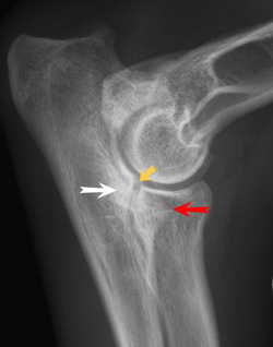
Elbow Dysplasia in DogsElbow dysplasia is a condition caused by the abnormal growth of cells, tissue, or bone. The condition is characterized by a series of four developmental abnormalities that lead to malformation and degeneration of the elbow joint. It is the most common cause of elbow pain and lameness, and one of the most common causes of forelimb lameness in large and giant-breed dogs. Labrador retrievers, Rottweilers, Golden retrievers, German shepherd dogs, Bernese mountain dogs, Chow Chows, bearded collies, and Newfoundland breeds are the most commonly affected. The age for onset of clinical signs is typically four to ten months, with diagnosis generally being made around 4 to 18 months.
One type of the condition is more likely to affect males than females: when the bone fragment is located at the inner surface of the upper ulna. The ulna is one of the bones of the foreleg, just below the elbow joint. Otherwise, there are no known gender differences.
Symptoms and Types
Causes
The causes are genetic, developmental, and nutritional.
Diagnosis
Your veterinarian will want to rule out several possible causes for the symptoms before arriving at a diagnosis. For example, whether there has been trauma to the joint, or whether there is an infection that has brought on, an arthritic condition will need to be explored. A tumor may account for the symptoms, and this possibility will be taken into account as well, with x-ray images taken of the affected area for closer examination. Both elbows will probably need to be x-rayed, since there is a high incidence of disease occurring in both legs. Your doctor may also want to order a computed tomography (CT) scan, or magnetic resonance image (MRI) to look for fragments. A sample of fluid will be taken from the joint with a fine needle aspirate for laboratory testing, and an arthroscopic examination (by use of a tubelike instrument for examining and treating the inside of the joint) may be utilized to help for making a definitive diagnosis.
Treatment
Surgery may be the treatment of choice; if so, cold-packing the elbow joint immediately following surgery to help decrease swelling and control pain is advised. You will want to continue to apply the cold pack five to ten minutes every eight hours for three to five days, or as directed by your veterinarian. Range-of-motion exercises will be beneficial for healing therapy until your dog can bear weight on the limb(s). Your veterinarian will demonstrate the types of range of motion movements you will be working on with your dog, based on the location and severity of the affected limb. Activity is restricted for all patients postoperatively for a minimum of four weeks, but to avoid muscle wasting or abnormal rigidity, you will need to encourage early, active movement of the affected joint(s). Again, have your veterinarian advise you on the specific movement therapy you will be using with your dog.
Weight control is an important aspect of decreasing load and stress on the affected joint(s). Medications may be prescribed for minimizing pain and decreasing inflammation. Medications may also be prescribed for slowing the progression of arthritic changes, and for protecting joint cartilage.
Prevention
Excessive intake of nutrients that promote rapid growth can have an influence on the development of elbow dysplasia; therefore, restricted weight gain and growth in young dogs that are at increased risk (due to breed, etc.) may decrease its incidence. Avoid breeding affected animals, since this is a genetic trait. If your dog has been diagnosed with elbow dysplasia, you will need to have it neutered or spayed, and you will need to report the incident to the breeder your dog came from, if that is the case. If the affected dog came from a litter in your own home, do not repeat dam–sirebreedings that result in these offspring.
Living and Management
Yearly examinations are recommended for assessing the progression and deterioration of joint cartilage. Progression of degenerative joint disease is to be expected; however, the prognosis is fair to good for all forms of this disease.
One type of the condition is more likely to affect males than females: when the bone fragment is located at the inner surface of the upper ulna. The ulna is one of the bones of the foreleg, just below the elbow joint. Otherwise, there are no known gender differences.
Symptoms and Types
- Not all affected dogs will show signs when young
- Sudden (acute) episode of elbow lameness due to advanced degenerative joint disease in a mature patient are common
- Intermittent or persistent forelimb lameness that is aggravated by exercise; progresses from stiffness, and noticed only after the dog has been resting
- Pain when extending or flexing the elbow
- Tendency for dogs to hold the affected limb away from the body
- Fluid build-up in the joint
- Grating of bone and joint with movement may be detected with advanced degenerative joint disease
- Diminished range of motion
Causes
The causes are genetic, developmental, and nutritional.
Diagnosis
Your veterinarian will want to rule out several possible causes for the symptoms before arriving at a diagnosis. For example, whether there has been trauma to the joint, or whether there is an infection that has brought on, an arthritic condition will need to be explored. A tumor may account for the symptoms, and this possibility will be taken into account as well, with x-ray images taken of the affected area for closer examination. Both elbows will probably need to be x-rayed, since there is a high incidence of disease occurring in both legs. Your doctor may also want to order a computed tomography (CT) scan, or magnetic resonance image (MRI) to look for fragments. A sample of fluid will be taken from the joint with a fine needle aspirate for laboratory testing, and an arthroscopic examination (by use of a tubelike instrument for examining and treating the inside of the joint) may be utilized to help for making a definitive diagnosis.
Treatment
Surgery may be the treatment of choice; if so, cold-packing the elbow joint immediately following surgery to help decrease swelling and control pain is advised. You will want to continue to apply the cold pack five to ten minutes every eight hours for three to five days, or as directed by your veterinarian. Range-of-motion exercises will be beneficial for healing therapy until your dog can bear weight on the limb(s). Your veterinarian will demonstrate the types of range of motion movements you will be working on with your dog, based on the location and severity of the affected limb. Activity is restricted for all patients postoperatively for a minimum of four weeks, but to avoid muscle wasting or abnormal rigidity, you will need to encourage early, active movement of the affected joint(s). Again, have your veterinarian advise you on the specific movement therapy you will be using with your dog.
Weight control is an important aspect of decreasing load and stress on the affected joint(s). Medications may be prescribed for minimizing pain and decreasing inflammation. Medications may also be prescribed for slowing the progression of arthritic changes, and for protecting joint cartilage.
Prevention
Excessive intake of nutrients that promote rapid growth can have an influence on the development of elbow dysplasia; therefore, restricted weight gain and growth in young dogs that are at increased risk (due to breed, etc.) may decrease its incidence. Avoid breeding affected animals, since this is a genetic trait. If your dog has been diagnosed with elbow dysplasia, you will need to have it neutered or spayed, and you will need to report the incident to the breeder your dog came from, if that is the case. If the affected dog came from a litter in your own home, do not repeat dam–sirebreedings that result in these offspring.
Living and Management
Yearly examinations are recommended for assessing the progression and deterioration of joint cartilage. Progression of degenerative joint disease is to be expected; however, the prognosis is fair to good for all forms of this disease.

 RSS Feed
